
Putting the “Eye” in CSI
Optical techniques help forensic investigators see crime-solving clues
Forensic investigation
hasn’t yet achieved the sci-fi type of technological sophistication seen on
CSI and other television crime shows, but it’s getting there. Having seen
what is possible with DNA, and often feeling considerable pressure to bring all
resources to bear – especially with high-profile casework – police institutions
are stepping up interest in a range of forensic technologies, including a number
of optical methods. This in turn is spurring development of new tools.
As a result, expect police departments and crime scene investigators
to become even more high tech than they already are, calling on a host of new techniques
for collection and analysis of evidence. Before we know it, they will look at least
a little bit more like what we see on TV.
Although technological advances are giving us ever more powerful
tools, there is no one silver bullet method that will meet all of an investigator’s
forensic needs. “Each application has a technique that is particularly suited
to it,” said Melanie Bailey, a research fellow at the University of Surrey’s
Ion Beam Centre in Guildford, UK. “In fact, it is normally useful to use a
number of techniques in complement to one another to get maximum information from
them.”
One example: Optical methods such as attenuated reflection Fourier
transform infrared (FTIR) spectroscopy offer up information about contaminants in
fingerprints – cosmetics, for example. But there may be ambiguities with respect
to the particular type of contaminant. For this reason, the investigator might conduct
a complementary analysis with secondary ion mass spectrometry or another similar
technique to confirm the nature of the contaminant detected.
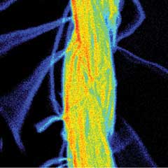
Particle-induced x-ray emission PIXE shows
promise for a range of forensic applications because it can be applied to a number
of types of evidence, including gunshot residue, glass fragments and paints. Shown
is a PIXE image of a fiber. Courtesy of Melanie Bailey, University of Surrey’s
Ion Beam Centre.
Still, a handful of technologies are showing particular promise
in forensics. Particle-induced x-ray emission (PIXE) can help to identify gunshot
residue from different sources, for instance. Finding such residue on an individual
might at first seem suspicious, but it could have come from the arresting officer
or from some other source. For this reason, it is important that investigators be
able to link the residue specifically to that found on the victim or at the crime
scene. Police currently use scanning electron microscopy to this end, but PIXE is
much more sensitive to small compositional differences in the residue. And the technique
can be applied to a range of other types of evidence, Bailey said, including soils,
glass fragments, fibers and paints.
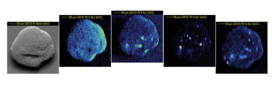 Optical techniques also can aid in soil analysis. Shown are elemental maps of quartz grains
from soil recovered from a forensic investigation. Courtesy of Melanie Bailey, University
of Surrey’s Ion Beam Centre.
Optical techniques also can aid in soil analysis. Shown are elemental maps of quartz grains
from soil recovered from a forensic investigation. Courtesy of Melanie Bailey, University
of Surrey’s Ion Beam Centre.
Advances in other areas also could contribute to analysis of such
residue – for example, in scanning electron microscopy/energy dispersive x-ray
spectroscopy, FTIR spectroscopy and matrix-assisted laser desorption/ionization
mass spectrometry, the latter of which has been shown to detect nicotine and other
contaminants in latent fingerprints.
Ambient MS: On-scene analysis
Among the many technologies available to them, police departments
and crime scene investigators are showing a keen interest in the rapidly developing
field of ambient mass spectrometry (MS). Encompassing the methods desorption electrospray
ionization (DESI), direct analysis in real time, plasma-assisted desorption ionization,
extractive electrospray ionization and others, ambient mass spectrometry enables
direct analysis of a wide range of substrates and thus shows promise in a number
of areas. These include toxicological studies, trace chemical residue analysis,
breath analysis, document verification and identification of counterfeits.
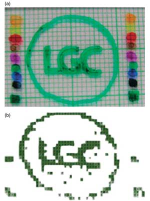
Desorption electrospray ionization mass spectrometry (DESI MS) can
contribute to a number of applications. Shown is an optical image of an ink LGC
Ltd. logo alongside a set of dyes for comparative identification, and a DESI MS
image of the molecular ion at 385 μm for the green pen molecular ion. Reprinted
with permission of Surface Interface Analysis.
Mass spectrometry generally allows investigators to determine
the chemical composition of a sample with high sensitivity. The mass spectral data
it provides can be compared with chemical databases, and it is this feature that
is critical to its use as evidence, said Felicia M. Green, a researcher with National
Physical Laboratory in Teddington, UK. She has worked extensively with ambient MS
techniques.
At the same time, the move toward the latter techniques is facilitating
direct analysis of surfaces without the pretreatment, filtering and extraction steps
traditionally needed in mass spectrometry. “This speeds up analysis and helps
to provide a simpler chain of custody to the results,” Green said.
Besides these benefits, the development of mini mass spectrometers
is contributing to on-scene chemical identification, both reducing sample turnaround
times and minimizing the possibility of contamination while transporting evidence
back to the lab. Liang Gao and co-workers at Purdue University in West Lafayette,
Ind., have reported a handheld, shoebox-size instrument, for example, that offers
parts-per-billion sensitivity. This and other systems have proved capable of detecting
cocaine on bank notes; explosives on paper, plastic and metal; and more –
all while sacrificing little in the way of performance.
Ambient MS methods still require some development before they
can be deployed in the field; for example, researchers will have to validate them
against results from the same technologies in different labs and against other technologies.
Also, Green said, “forensic data must stand up to scrutiny in a court of law,
so it is essential that a robust measurement infrastructure is developed to support
accredited measurements using accepted quality systems.” To achieve this,
methods must prove capable of providing repeatable, reproducible and valid results.
In addition, the construction of databases is critical, so researchers
can ascertain the significance of the results. Here, Bailey said, the overwhelming
obstacle appears to be the difficulty in obtaining funding. “This requires
considerable investment. Funding councils are not interested in database construction,
since this does not further science in any way in their opinion. However, without
funding for such activities, it is a real challenge to demonstrate the usefulness
of the techniques to police institutions.”
CT and forensic radiology
Although recent decades have seen significant advances in clinical
radiology, forensic application of imaging methods has not always kept pace. Conventional
x-ray often will make an appearance, but newer technologies such as computed tomography
(CT) are still not as widely used as they could be in forensic investigations.
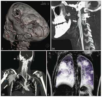
Investigators have shown that computed tomography (CT) can provide an important complement to
traditional autopsies. Here, a volume-rendering CT technique clearly depicts a transverse
fracture line at the base of the skull in a car crash victim. Reprinted with permission
of Archives of Pathology & Laboratory Medicine.
Postmortem investigations in particular could benefit from application
of these methods. CT and other similar techniques could provide an objective and
nondestructive alternative to – or at least complement – the traditional
autopsy. So, for example, if new evidence were to arise, the investigator could
return to the 3-D scan performed prior to the autopsy to seek new understandings
based on that evidence. Otherwise, he or she would likely have to rely on written
documentation or specimens kept for histological examination.
Also, the techniques can be used in cases where grieving relatives
do not want an autopsy performed, or they can provide visual demonstrations when
needed in the courtroom.
Investigators have been exploring the potential of both multislice
computed tomography and whole-body computed tomography angiography. Patricia M.
Flach and colleagues at the University of Bern in Switzerland reported on the latter
in the January 2010 issue of Archives of Pathology & Laboratory Medicine. Applying
it in the case of a fatal car crash, they were able to detect multiple injuries
of the organs in the upper abdomen as well as of lesions of the brain parenchyma
and vasculature of the neck.
The CT findings agreed completely with those of traditional autopsy
and even aided interpretation in areas that can be difficult to access through manual
dissection.
Published: September 2010