By Lynn Savage, Photonics Spectra Features Editor
Quantum dots, also known as semiconductor artificial atoms, have existed for several years now, and their use keeps on expanding. Primarily used as substitutes for dyes in fluorescence imaging, their propensity for being highly tunable emitters that resist photobleaching represents only a small fraction of their benefits.
Commonly but not always comprising cores of cadmium selenide with a coating of zinc sulfide, quantum dots are perhaps best known for the tunability of their emission wavelength, once excited by an external light source: Change the size of the particle, get a specific wavelength in return. Early research into the properties and potential uses for quantum dots kept them in solutions, and then later brought into proximity with a variety of other particles. This led to their nearly ubiquitous use as donors in Förster's resonance energy transfer (FRET), a renowned method for near-diffraction-limit imaging of cells and other subjects.
Scientists, however, aren't keeping still, and are looking for ways to improve quantum dots as an imaging resource as well as to expand their roles into new areas altogether.
Watching transfection
Gene therapy is tricky work, but with tremendous potential for treating cancer and other diseases. The base idea is to gain control over the mechanism of cells so that they can, for example, resist cancer, destroy tumors or make tumors more susceptible to chemotherapy. Delivering specially designed genetic material into cells — via transfection — is a delicate process that involves carrying DNA or RNA fragments to a cell, protecting them from external forces, and then broaching the cell membrane and releasing the payload. Once released from its polymeric vessel, or vector, the genetic material alters the expression of genes with a cell, thereby changing the cell's function.
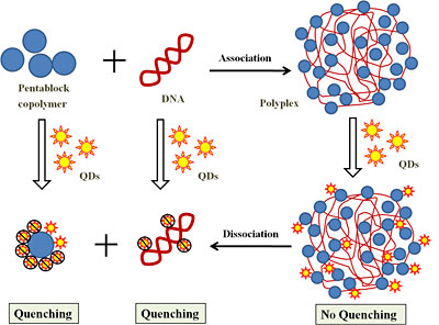
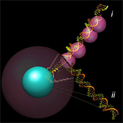
Researchers at Iowa State University have found that quantum dots can be used to measure the dissociation of DNA/polymer complexes within cells, an important step toward reliable cancer treatment via gene transfection. Shown is a schematic of the mechanism by which quantum dot fluorescence reveals the dissociation of DNA from pentablock copolymers. (Reused with permission of American Chemical Society)
There are numerous ways to deliver genetic material to cells (a process known as transfection), but until recently no sure way of measuring how well one's attempt succeeded. One way is to use fluorescence microscopy to measure the distance between payload and carrier while they dissociate inside a cell.
Quantum dots, which are frequently used as substitutes for chemical dyes in fluorescence microscopy, are used by Aaron R. Clapp and his colleagues at Iowa State University in Ames. In the Jan. 25, 2011, issue of ACS Nano, Clapp's group reported its efforts to determine how well quantum dots are suited to sensing the dissociation of DNA payloads held within complexes of pentablock copolymers.
In some approaches to gene therapy, Clapp said, freeing the genetic payload from its delivery vehicle represents a significant hurdle to efficient gene expression. The group's purpose, then, was to devise a method for directly measuring or observing the dissociation process between DNA and the pentablock copolymer carrier.
"There are often conflicting requirements during each stage of gene delivery where the DNA must be tightly bound and protected by the vector, but later released once delivered inside the cell," he said. "Each of these steps represents a potential barrier to gene delivery, and the overall efficiency is limited by the least efficient step in the process."
The team capped CdSe-core/ZnS-shell quantum dots with the amino acid cysteine to improve their water-solubility and the ligand-exchanging process. The capped quantum dots also have a net negative charge, which increased bonding with the positively charged pentablock copolymers. After adding DNA fragments to the quantum dot/polymer complex, the researchers introduced it to tumor cells then added chloroquine, an agent known to promote dissociation.
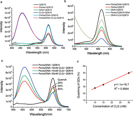
(a-c) show the effects of DNA, chloroquine (CLQ) and pentablock copolymer (penta) — alone or in combination — on the fluorescence of quantum dots that have an emission maximum of 615 nm (QD615). (d) plots QD615 quenching versus chloroquine concentration. (Reused with permission of American Chemical Society)
Using either a xenon light source or an argon-ion laser emitting at 488 or 514 nm, they excited the quantum dots and measured the amount of light from the semiconductor particles. Whereas chloroquine alone had no effect on the emissions of quantum dots, eliciting dissociation of the DNA/polymer/quantum dot complex with chloroquine caused significant quenching compared with control samples to which chloroquine was not added.
The results "suggest that quantum dots are good candidates for elucidating the complex process of delivery and release within cells," Clapp said. "This provides a useful technique for quantifying release of genetic cargo and visualizing the process using fluorescence microscopy."
Analyzing hybridization
Using quantum dots as the basis of biosensors is increasing. In another such use, Ulrich J. Krull and his colleagues at the University of Toronto, Mississauga, are constructing hybridization assays that may bring a new level of portability to human and veterinary medical diagnoses as well as to the detection of food pathogens at their sources.
In a paper published by Analytical and Bioanalytical Chemistry in January 2011, Krull and his team describe efforts to create a solid-phase nucleic acid hybridization assay in which CdSe-core/ZnS-shell quantum dots are immobilized within the walls of microfluidic channels.
Detection of solid-phase targets is often accomplished with microarrays comprising hundreds of tiny pads composed of unique, single-stranded probe sequences of DNA or RNA. Passing a sample over these arrays can show how gene expression is triggered by any of the probes. Such microarrays and another common method of detecting gene expression — real-time polymerase chain reaction — can be expensive, unwieldy and too free with the required chemical components, however.

Quantum dots immobilized onto the inner walls of a microfluidic system's channels acts as FRET donors, thus completing a solid-phase DNA hybridization assay that could result one day in a portable pathogen detector. The quantum dots are arranged at a density of 1012 to 1013 per square centimeter. (Courtesy of Ulrich Krull, University of Toronto, Mississauga)
"These concerns represent an opportunity for biosensors," Krull said, "devices that immobilize probe material directly to a transduction element that translates the biorecognition event into a measurable analytic signal."
Krull and his colleagues use quantum dots that are coated with fluorescent nucleic acid probes. The quantum dots serve as donors in FRET events when hybridization of the target molecules and the nucleic acids probes occurs.
Hybridizing oligonucleotides onto quantum dots reduces interference and improves sensitivity of the FRET process during experiment. (Courtesy of Ulrich Krull, University of Toronto, Mississauga)
"Different DNA probe sequences can be associated with different-sized quantum dots to produce a range of unique biosensors that are distinguished using emission wavelength channels," Krull said. Thus, using a single excitation source - in this case, 405-nm laser from Coherent Inc. - Krull's group can elicit multicolor emissions, with each color signifying a different potential pathogen or other target.
Immobilizing the quantum dot and DNA probes to the inner walls of a microchannel system enables the use of small sample volumes and, overall, permits shrinking of the entire assay to an easily portable footprint. The goal, Krull said, is to put the quantum dot based bioprobe microchannel system onto the surface of a CMOS chip. Other advantages include an increase in the speed of signal generation from 15 to 20 min for microarray technologies and RT-PCR to just a few minutes or even seconds and regeneration, so that a system does not need disassembling or new cartridges added when trying to accomplish higher-throughput analyses.
"The spectral imaging system will be simplified by exciting the quantum dots directly on top of a spectrally sensitive photodetector fabricated on a CMOS microchip, thereby creating a sensitive, low-cost integrated detection system," he said.
The chemical truth
As seen in the previous examples, quantum dots are no longer allowed to roam free, nor are they even just loosely confined. Instead, they typically are conjugated with one sort of (bio)molecule or another, with the goal of improving the view of whatever target they are helping to shed light on. However, it is possible that many researchers are taking quantum dot conjugation for granted.
"We realized a long time ago that in order to get the hybrid quantum dot bioconjugates to function exactly as we intended for a given sensing or imaging application, we really need to have access to highly controlled bioconjugation chemistries and a very good understanding of the physical characteristics and subsequent activity of a given quantum dot bioconjugate," said Igor I. Medintz, a research biologist at the U.S. Naval Research Laboratory's Center for Bio/Molecular Science and Engineering in Washington. He and his group have been working with the semiconductor particles for about nine years.
Scientists at the Naval Research Laboratory are concerned that not enough is known about how the chemistry used to create bioconjugates has on their final structure. (Reused with permission of American Chemical Society)
Medintz and his colleagues at the center along with the Division of Optical Sciences and the Scripps Research Institute in La Jolla, Calif., study the way in which various peptides, proteins and nucleotides attach themselves to quantum dots when forming conjugates.
"When most people assemble a nanoparticle bioconjugate, they likely picture its composite structure in a somewhat idealized manner," Medintz said. "For example, conjugates comprising nanoparticles and antibodies generally are assumed to exist as a central nanoparticle surrounded symmetrically by a known number of antibodies. Furthermore, each antibody is attached to its nanoparticle in the same way - with its binding site facing outward and clearly available."
"This is the nanoparticle-bioconjugate that people want and expect from the chemistry," he said. "The reality is much different."
For example, in the above scenario, some nanoparticles may be crosslinked with one another, as might be some of the antibodies.
In the Nov. 17, 2010, online edition of ACSNano, Medintz's group took a close look at the attachment chemistry behind quantum dot bioconjugates. To do so, they used two separate common self-assembly techniques to attach DNA to quantum dots, and measured the resulting structures and compared them with predicted structures.
One conjugation type - modifying the DNA to display a polyhistidine sequence - yielded conjugates as predicted. Furthermore, FRET measurements of dye-labeled DNA hybridized to the DNA attached to the quantum dots matched expectations fairly well. Conjugates made using DNA capped with biotin and assembled to streptavidin-coated quantum dots, however, displayed good FRET qualities but failed to match expected FRET separation distances, indicating they assembled into unexpected structures.
The group concluded that the attachment chemistry used to assemble quantum dot/DNA conjugates has a strong influence on the conjugates' final structure, which in turn affects their functionality.
In addition to a better understanding of the chemistry involved, Medintz said, better tools are needed going forward for characterization and analysis.
"I am continually surprised by what bioconjugated quantum dots are capable of and how many of their intrinsic properties are almost untapped."
Lynn Savage
Photonics Spectra Features Editor