Researchers from the University of Bordeaux have found a way to calibrate stimulated emission depletion (STED) microscopy for improved brain imaging. The method increases the depth at which STED microscopy can image and be applied to other tissues.
STED microscopy requires the target sample to contain fluorophores — compounds that absorb light at one wavelength and then reemit it at a longer one. In the simplest version of the technique, fluorophores are excited in a circular spot by irradiation with a diffraction-limited focused laser. Then, a doughnut-shaped portion around the spot is irradiated with less-energetic light, called the depletion beam, that switches off the fluorescence by the process of stimulated emission.
The net effect is that only the fluorophores in the center of the doughnut reemit photons. Because that area can be made arbitrarily small, this allows for superresolution microscopy.
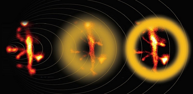
Multiphoton STED microscopy enhanced by adaptive optics captures the fine details of neuronal dendrites. Courtesy of Bancelin et al.
However, when imaging a tissue sample at depths greater than 40 μm, the depletion beam suffers various types of defocusing and degradation (or aberration). The researchers targeted the most common and troublesome type: spherical aberration.
The Bordeaux team prepared a brain tissue phantom sample, a gel-based substitute with a similar refractive index to that of the actual brain. The phantom sample contained homogenously dispersed fluorophores and gold nanoparticles, which allowed the team to clearly visualize and quantify how the shape of the depletion beam distorted as it traveled deeper into the sample. With this data, the team calculated necessary pre-adjustments to be applied to the depletion beam according to the tissue depth so that the final shape more closely matched the ideal one.
To apply these adjustments, the team used adaptive optics, a technology originally devised by astronomers to improve telescopic images suffering from aberrations caused by Earth’s atmosphere.
With the shape of the depletion beam calibrated, the team imaged live neural tissue. The results were compared against uncorrected STED microscopy and two-photon microscopy, which is a technique specifically adjusted for deep tissue imaging.
“Using our calibration strategy, we could measure neuronal structures as small as 80 nm at a depth of 90 μm inside biological tissue and obtain a 60% signal increase after correction for spherical aberration,” said Valentin Nägerl, a professor at the University of Bordeaux.
“Superresolution microscopy has been primarily applied for thin specimens, such as single-layer cells where light scattering is negligible,” said Ji Yi, a professor of biomedical engineering at Johns Hopkins University. “The team led by Valentin Nägerl implemented adaptive optics in a two-photon stimulated emission depletion microscopy, and achieved 80-nm resolution of imaging neuron dendritic spines through 90-μm brain tissue. This is noteworthy because superresolution is hard to maintain in thicker tissue, particularly given the highly scattering quality of brain tissue.”
The calibration process could be incorporated into standard laboratory practices to obtain better results with STED microscopes, provided the prepared phantom sample matches the optical properties of the biological specimen.
“Our approach is not limited to brain samples; it could be adapted to other tissues with known and relatively homogenous refractive indices, as well as other types of preparations, even potentially in the intact, live mouse brain,” Nägerl said.
The research was published in Neurophotonics (www.doi.org/10.1117/1.NPh.8.3.035001).