
Scalpel-free surgery with deep-tissue imaging
A new procedure that more than doubles the depth that light can be focused inside biological tissues soon could enable doctors to perform incision-free surgery or diagnose cancer by seeing tumors inside the body.
Although the previous limit for how deep light could be focused into tissue was only about 1 mm, researchers at California Institute of Technology (Caltech) now can reach 2.5 mm. In principle, their method could focus light as much as a few inches into tissue.
“The ability to focus high-intensity light tightly deep within tissue has a lot of applications,” said Changhuei Yang, a professor of electrical engineering and bioengineering at Caltech. “We hope that with further technology improvement, depth up to a few centimeters will be achievable. If we can reach up to about 10 cm, it would allow us to reach most regions of the human anatomy.”
The new technique builds on a previous method that Yang and his colleagues developed to see through a layer of biological tissue, which is opaque because it scatters light. In that study, the scientists shone light through a tissue sample and recorded the resulting scattered light on a holographic plate. The recording contained information about how the light beam scattered, zigzagging through the tissue. By playing the recording in reverse, they sent the light back through the other side of the tissue, retracing the beam’s path to the original source.
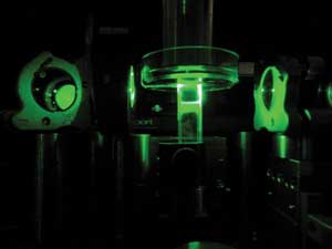
A new technique more than doubles the depth that light can be focused inside biological tissue. In the experiment, Caltech researchers shone green laser light into the tissue sample seen here in the center.Images courtesy of Caltech/Benjamin Judkewitz and Ying Min Wang.
In this way, they could send light through a layer of tissue without the blurring effect of scattering. However, to make images of the tissue’s insides, they would have to be able to focus a beam of light into it.
To precisely focus light into tissue, the team expanded upon the recent work of Lihong Wang’s group at Washington University in St. Louis (WUSTL), which developed a technique to focus light using the high-frequency vibrations of ultrasound and two of ultrasound’s favorable properties. First, its high-frequency sound waves are not scattered by tissue; second, its ultrasonic vibrations interact with light in such a way that the light’s frequency is shifted ever so slightly. As a result of this acousto-optic effect, light that interacts with ultrasound changes into a slightly different color.
Both teams focused ultrasound waves into a small region inside a tissue sample during their experiments. Next, they shone light into the sample, which scattered the light. Any light that passed through the region with the focused ultrasound changed color somewhat. The researchers identified and recorded the color-shifted light.
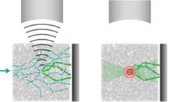
Left, light enters the tissue sample and is scattered (blue arrows). From above, ultrasound is focused into a small area inside the tissue. The ultrasound shifts the frequency of any light that passes through that area ever so slightly, changing its color. The color-shifted light (green) is then recorded. Right, the recorded light is sent back to retrace its steps to the small region where the ultrasound was focused – which means the light itself is focused on that area.
Using Caltech’s playback technique, they sent the light back, inducing only the color-shifted portion to retrace the path to the small region where the ultrasound was focused. This means that the light itself is focused on that area, allowing an image to be created. By moving the ultrasound’s focus, the researchers can control where they want to focus the light.
Only a very small amount of light could be focused in the WUSTL experiment, but Caltech’s method allows scientists to fire a beam of light with as much power as they need for potential applications.
“This technology is still in its infancy,” Yang told BioPhotonics. “We took an important step beyond Lihong Wang’s original demonstration of TRUE (time-reversed ultrasonically encoded optical focusing) by implementing a TRUE technique that is effectively unlimited in terms of its ability to deliver arbitrarily high power to the focused spot.”
For this to work in living tissue, Yang said, the team must decrease the time for generating a focused light spot to a fraction of a second, depending on the tissue type. “The ability to build a suitable system is within our technological reach. But it does require a significant financial investment to make it happen. If we have the financial resources and a semiconductor foundry to help us, bringing the technology to the point of clinical imaging is like a 10- to 20-year process.”
The team demonstrated how the new method could be used with fluorescence imaging by embedding a patch of gel with a fluorescent pattern that spelled out “CIT” inside a tissue sample. The investigators scanned the sample with focused light beams, which hit and excited the fluorescent pattern, resulting in the glowing letters emanating from inside the tissue. They also used the technique to take images of tumors tagged with fluorescent dyes.
The fluorescence is used only during the final imaging scan, Yang said, and the technique works whether the dye is used or not.
“We can form a focused light spot [regardless],” he said. “We chose to do fluorescence imaging here because our technique is able to generate a sufficiently strong focused spot to excite fluorophores to provide image contrast.”
Doctors also could use the technique to treat cancer with photodynamic therapy, which currently can be used only at the surface of tissue because of the way light is easily scattered. The new method should make it possible to reach cancer cells deeper inside tissue.
Next, Yang said he and his team would like to combine the spatial light modulator and image sensor into a single digital optical phase conjugation chip, but funding is a challenge. If they could achieve this single chip, Yang said, it would “solve several technical implementation challenges in one move and, more importantly, allow us to deploy sensing and playback over a far larger area. The more area we can cover, the task of collecting and playing back the conjugate light field becomes faster and easier.”
The study appeared in Nature Communications (doi:10.1038/ncomms1925).
Published: September 2012