Dual-camera approach images expansion and contraction of microdroplets as they are hit by acoustic waves.
Frank Kosel, IMC/Photo-Sonics Inc.
The ability to record framing and streak images simultaneously has long been a requirement in the research fields of electrical discharge, biomedical research and other applications. Early systems incorporating framing and streak cameras were large and cumbersome, using two optical paths or low-efficiency beamsplitting optics.
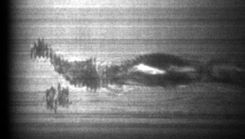
A streak image shows from left to right a total time of 20 μs and a slit width of 50 μm, providing 50-ns time resolution. The droplet diameters are shown as straight lines at the left of the image before an acoustic wave hits the droplets. As the acoustic wave arrives, the droplet diameter expands and contracts in a matter of nanoseconds. The dark area that turns to light at the start of the streak shows the turn-on time of the flashlamp.
Capturing framing and streak images of ultrafast phenomena, such as microdroplets being struck by acoustic waves, simultaneously remains a technical challenge. Researchers wanted to record these images through a microscope at 20× to 200× at speeds of 1 million fps and at a streak-time resolution of 200 ns over a 40-μs time window with a portable system.
A new system, built by IMC/Photo-Sonics Inc. of Burbank, Calif., and delivered to Oliver D. Kripfgans, an assistant professor in the department of radiology at the University of Michigan in Ann Arbor, incorporated an SIM02-16 ultrafast framing camera manufactured by Specialised Imaging Ltd. of Tring, UK, and an Optoscope SC-10 streak camera manufactured by Optronis GmbH of Kehl, Germany, into a single package that attained performance levels never before achieved with a dual-camera system.
Recording the cycles of contraction and expansion of microdroplets and bubbles in a liquid medium, when hit with an acoustic shock wave, has been of keen interest to Kripfgans for many years. The size and speeds at which these events take place make imaging a challenge. An acoustic wave in liquid travels at ∼1500 m/s. Transition time through microdroplets is on the order of tens of nanoseconds. Cyclical events in the droplet occur in submicroseconds and longer. Thus, to stop the motion, there is a need for speeds of >1 million fps and for exposure time resolution of less than 100 ns.
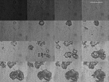
In this image, the framing rate is 3.3 million per second and the exposure time per frame, 50 ns, for a total recording time of 4.55 μs. The frame sequence goes from left to right and from top to bottom. Frame 4 shows the relative position of the slit placed over the diameter of the droplets for the streak camera image. The acoustic wave arrives between frames 5 and 6, and the droplets start to expand over the next 4.5 μs. The dark frame at start shows the turn-on time of the flashlamp.
A newly designed optical beamsplitter — which is optimized for >70 line pairs per mm spatial resolution and for no geometric distortions and which can be configured with up to 16 output ports — is the primary imaging system. The splitter used for this test had nine optical ports, eight of which were fitted with intensified charge-coupled devices (ICCDs) for framing operation. The ninth port allowed a full-size streak camera to be coupled. Having the streak camera external but integral to the imaging system allowed the use of a large-format streak camera (with a 16-mm-long slit and a 20-mm screen), ensuring maximum spatial resolution, long record times and access to the input streak optics.
The framing camera has 16 frames and houses the beamsplitter fitted with eight ICCDs, each producing two frames of 1360 × 1024 pixels at 12 bits analog-to-digital (A/D) per frame. The speeds are programmable from 100 fps to 200 million fps with exposure times from 10 ms to 5 ns. Adjustable gain from unity to ∼10,000× as well as multiple exposures are possible. Pixel size is 6.45 μm square.
For these tests, the framing camera was programmed for 3.3 million fps and 50-ns exposure times, providing a total recording time of 4.55 μs. The trigger input and output pulses allow for synchronization to and from other devices, such as pulsed lasers or flashlamps, and can be used to verify critical timing with respect to other events associated with the experiment.
The streak camera, meanwhile, is built around the 2-ps time-resolution streak tube with an 8-mm photocathode and a 20-mm screen. It has programmable sweep speeds from 330 ps/mm to 5 ms/mm and includes a removable 25-mm microchannel plate intensifier and a fiber optic-coupled CCD camera to capture and digitize the streak data with 1392 x 1024 pixels at 12 bits A/D per image.
The streak camera was programmed for 1 μs/mm, providing a 20-μs time window. Using a 50-μm slit gave 50-ns time resolution, equaling the exposure time of the framing camera but with a much longer recording time. The two-to-one magnification input streak optics allowed recording a 16-mm slit image of the event.
The two cameras were mounted on a rigid plate and run in live-focus mode. This setup allowed critical focusing and alignment through the video viewing port on the inverted microscope. Microdroplets ranging from 10 to 50 μm in diameter in water were positioned into the field of view of the microscope, which was run at 40× magnification and which gave 0.163-μm-per-pixel resolution at the field of view.
An acoustic pulse generator was used to send a high-energy acoustic wave to the event and a synchronized pulse to the cameras. The time of arrival of the acoustic wave to the droplet was used as the initial delay to the start of the recording sequence. An IMS-300 point xenon flashlamp that provided 300-J, 25-μs pulses was fitted with relay optics and a fiber optic lightguide. It was sent a prepulse 10 μs ahead to ensure peak intensity to backlight the droplets.
Test shots verified trigger timing, lighting and alignment. With all of the parameters set, a data shot was taken by applying an acoustic pulse to the water bath. As the shock wave passed through the droplets, a sequence of frames and a streak record showed droplet deformation as a function of time and space.
Framing images show extremely detailed two-dimensional information about the full field of view, whereas the streak image is a continuous chronological record of a slit image of the diameter of the droplets. Together, the cameras provide a unique insight into the visualization of nano events.
Meet the author
Frank Kosel is senior sales and applications engineer of the ultrahigh-speed imaging systems at IMC/Photo-Sonics Inc. in Burbank, Calif.; e-mail: [email protected].