
Single Microendoscope Acquires Photoacoustic, Fluorescent Images
An endoscope that combines photoacoustic and fluorescence imaging and that is about as thick as a human hair could one day provide insight into the brain by enabling the simultaneous measurement of blood dynamics and neuronal activity. The researchers responsible for developing the device fabricated a prototype microendoscope that measured just 250 × 125 µm2, and used it to image fluorescent beads and blood cells using both imaging modalities. They successfully detected multiple 1-µm fluorescent beads and individual 6-µm red blood cells.
“Combining these imaging modalities could improve our understanding of the brain’s structure and behavior in specific conditions such as after treatment with a targeted drug,” said research team leader Emmanuel Bossy from the CNRS/Université Grenoble Alpes Laboratoire Interdisciplinaire de Physique. “The endoscope’s small size helps minimize damage to tissue when inserting it into the brains of small animals for imaging.”
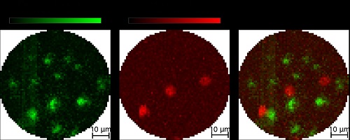
The researchers demonstrated their new device by using it to image fluorescent beads (green) and red blood cells. The FOV is the size of a hair. Courtesy of Emmanuel Bossy, CNRS/ Université Grenobe Alpes Laboratoire Interdisciplinaire de Physique.
The device enables the automatic co-registering of fluorescence and photoacoustic images with complementary information. Fluorescent signals, which are created when a fluorescent marker absorbs light and reemits it with a different wavelength, are useful for labeling specific regions of tissue. Conversely, photoacoustic images, which capture an acoustic wave generated after the absorption of light, do not require labels, and this can be used to image blood dynamics, for example.
The new endoscope uses a technique called optical wavefront shaping to create a focused spot of light at the imaging tip of a very small multimode optical fiber.
“Light propagating into a multimode fiber is scrambled, making it impossible to see through the fiber,” Bossy said. “However, this type of fiber is advantageous for endoscopy because it is extremely small compared to the bundles of imaging fibers used for many medical endoscopic devices.”
A spatial light modulator sends specific light patterns through the fiber and creates a focus spot at the imaging end. When the focus spot hits the sample, it creates a signal that can be used to build up an image point-by-point by raster scanning the spot over the sample.
To add photoacoustic imaging to the endoscope, the researchers incorporated a thin optical fiber with a sensor tip sensitive to sound. As commercially available fiber optic acoustic sensors aren’t sensitive or small enough for the application, the researchers turned to a fiber optic sensor developed by Paul Beard of the University College of London’s research team.
“The focused spot of light allows us to build the image pixel by pixel while also increasing the strength of fluorescence and photoacoustic signals because it concentrates the light at the focal spot,” Bossy said. “This concentrated light combined with a sensitive detector made it possible to obtain images using only one laser pulse per pixel, whereas commercial fiber optic acoustic sensors would have required many laser pulses.”
The researchers are currently working to increase the device’s acquisition speed, with the goal of acquiring multiple images per second.
The study was published in Biomedical Optics Express (www.doi.org/10.1364/BOE.400686).
Published: September 2020