Search
Menu
Trioptics GmbH - Worldwide Benchmark 4-24 LB

Technique Calibrates Hyperspectral Sensors

Facebook X LinkedIn Email
Software that calibrates hyperspectral imagers could ensure that their performance is optimized for medical applications, such as determining the diseased tissue margins during surgery.

Because of their superior ability to identify objects by color (they can distinguish the full color spectrum in each pixel) and because diseased tissues and cells have distinct spectral signatures, hyperspectral imagers (HSIs) are being pursued for use in medical applications. The imaging technique is currently used in remote sensing.

“Because diseased tissues and cells also have distinct spectra, scientists have been trying to use HSI for medical applications as well,” said National Institute of Standards and Technology physicist Jeeseong Hwang. “But anytime you tell a machine to scan for something, you need to be sure it is actually looking for what you want, and you have to make sure that the image analysis algorithm extracts the correct color information out of a complex multicolor data set. We decided to create a way to calibrate an HSI device and to test its algorithm as well.”


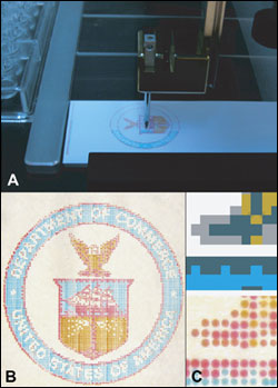
(a) Microarrayer machines now can mix colors and deposit them on microscope slides, which can be used to calibrate hyperspectral imagers (HSIs) used in medical applications. (b) The finished slides can be custom-colored to calibrate HSIs to find specific types of tumors or diseased tissue. (c) Close up, they resemble dot-matrix print work. (Image: Clarke/NIST)


Vescent Photonics LLC - Lasers, Combs, Controls 4/15-5/15 MR
New software was developed by Matthew Clarke, a former National Research Council-supported postdoctoral fellow in Hwang’s group who now is working in the National Gallery of Art in Washington, for a microarrayer device, which was named as such because it can lay down hundreds of tiny sample droplets in specific places on a microscope slide’s surface. Microarrayers typically create DNA arrays for genetic research, but the NIST team remade it into an artistic tool by programming it to select chemicals of different hues and lay them down on the surface of a slide. 

The results, which look like dot-matrix printing, can be used to calibrate medical HSI devices and image analysis algorithms. This could help surgeons better detect cells with a specific chemical makeup, which is determined by the cells’ color.

“Scientists and engineers can create a custom slide with the exact colors representing the chemical makeup they want the HSI devices to detect,” Hwang said. “It could be a good way to make sure the HSI devices for medical imaging perform correctly so that surgeons are able to see all of a tumor or diseased tissue when operating on a patient.”

The project is part of a larger NIST effort to evaluate and validate optical medical imaging devices.

The findings appeared in a special issue of Biomedical Optics Express.

For more information, visit: www.nist.gov

Published: June 2012
Glossary
hyperspectral imaging
Hyperspectral imaging is an advanced imaging technique that captures and processes information from across the electromagnetic spectrum. Unlike traditional imaging systems that record only a few spectral bands (such as red, green, and blue in visible light), hyperspectral imaging collects data in numerous contiguous bands, covering a wide range of wavelengths. This extended spectral coverage enables detailed analysis and characterization of materials based on their spectral signatures. Key...
microscope
An instrument consisting essentially of a tube 160 mm long, with an objective lens at the distant end and an eyepiece at the near end. The objective forms a real aerial image of the object in the focal plane of the eyepiece where it is observed by the eye. The overall magnifying power is equal to the linear magnification of the objective multiplied by the magnifying power of the eyepiece. The eyepiece can be replaced by a film to photograph the primary image, or a positive or negative relay...
photonics
The technology of generating and harnessing light and other forms of radiant energy whose quantum unit is the photon. The science includes light emission, transmission, deflection, amplification and detection by optical components and instruments, lasers and other light sources, fiber optics, electro-optical instrumentation, related hardware and electronics, and sophisticated systems. The range of applications of photonics extends from energy generation to detection to communications and...
remote sensing
Remote sensing is a method of data collection and observation where information about objects, areas, or phenomena on Earth's surface is gathered from a distance, typically using sensors onboard satellites, aircraft, drones, or other platforms. This technique enables the monitoring and analysis of Earth's surface and atmosphere without direct physical contact. Remote sensing systems capture electromagnetic radiation (such as visible light, infrared, microwave, or radio waves) reflected or...
AmericasBiophotonicscell detectionchemicalsdiseased tissueDNA arraysHSIhyperspectral imager calibrationhyperspectral imagershyperspectral imagingImagingJeeseong HwangMarylandMatthew Clarkemedical applicationsmicroarrayermicroscopeMicroscope SlideMicroscopyNational Gallery of ArtNational Research CouncilNISToptical medical imagingphotonicsremote sensingResearch & Technologyspectral signaturesTest & Measurement

We use cookies to improve user experience and analyze our website traffic as stated in our Privacy Policy. By using this website, you agree to the use of cookies unless you have disabled them.