Researchers from Tokushima University have developed a method of fluorescence lifetime microscopy that does not require mechanical scanning. The technique drastically increases the effectiveness of fluorescence lifetime microscopy (FLIM).
A major limitation to traditional FLIM methods stems from the fact that fluorescence decay is fast-occurring — most ordinary cameras are unable to capture it. Using a single-point photodetector overcomes the issue, though the device must be scanned over the area of a sample to construct an image with each measured point. The process involves the movement of mechanical pieces, which limits the speed of image capture.
According to Takeshi Yasui, a professor at the Institute of Post-LED Photonics at Tokushima University, “Our method can be interpreted as simultaneously mapping 44,400 light-based ‘stopwatches’ over a 2D space to measure fluorescence lifetimes — all in a single shot and without scanning.”
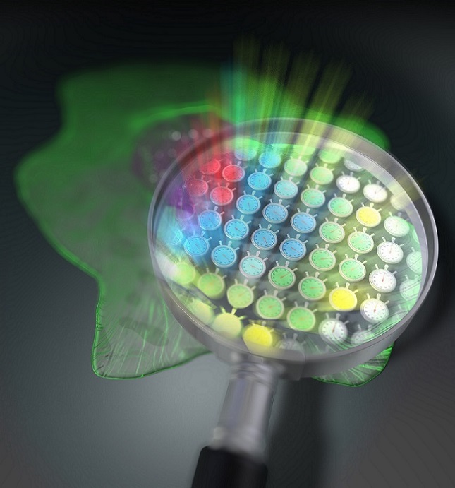
A visual representation of the fluorescence lifetime imaging technique developed at Tokushima University. Courtesy of Tokushima University.
The method uses an optical frequency comb as the excitation light for the sample; optical frequency combs are light signals composed of the sum of many discrete optical frequencies with a constant spacing between them. How the signal looks when plotted against optical frequency pertains to the “comb”: a tightly confined cluster of equidistant spikes extending from the optical frequency axis.

With special optical equipment, a pair of excitation frequency comb signals decomposes into individual optical beat signals (dual-comb optical beats). These feature distinct intensity-modulation frequencies, each of which carries a single modulation frequency and are irradiated onto a sample. Each light beam hits the sample in a unique location, resulting in a one-to-one correspondence between each point, or pixel, on the 2D surface of the sample and each modulation frequency of the dual-comb optical beats.
Due to its fluorescence properties, the sample then reemits part of the captured radiation while preserving the frequency-position correspondence. The fluorescence emitted from the sample is then focused with a lens onto a high-speed single-point photodetector, and the measured signal is able to appear in the frequency domain.
From the relative phase delay between the excitation signal at that modulation frequency versus the one measured fluorescence, the researchers were able to calculate each pixel.
Due to its superior speed and high spatial resolution, the new method simplifies the ability for users to apply the advantages of fluorescence lifetime measurements, which are of particular benefit in live cell imaging.
“Because our technique does not require scanning, a simultaneous measurement over the entire sample is guaranteed in each shot,” Yasui said. “This will be helpful in life sciences where dynamic observations of living cells are needed.”
Additionally, the approach could be used for the simultaneous imaging of multiple samples for antigen testing, which is already being used for the diagnosis of COVID-19.
The research was published in Science Advances (www.doi.org/10.1126/sciadv.abd2102).