Optical coherence tomography (OCT) imaging has been used primarily for ophthalmology to diagnose and treat retinal diseases such as glaucoma. In this arena, the technology is advancing rapidly with continuous R&D developments.
And while ophthalmology continues to dominate OCT, the landscape is expanding. It’s finding application in dermatology for diagnosis and treatment of skin cancers, and it is even guiding some laser-based aesthetic treatments such as skin rejuvenation.
OCT angioplasty (OCT-A) — derived from algorithms originally created for existing OCT technologies — is among promising breakthroughs, as it allows noninvasive imaging without the need for dyes and can produce images of blood flow in just a few seconds. Expanded field OCT offers high-speed imaging, producing faster data collection for clinical management. OCT is also being paired with confocal microscopy in some cases, as it offers deeper imaging compared to confocal alone, which has a limited depth of focus.
Several OCT experts recently talked with BioPhotonics, offering insight into how advancements in this field could restructure the future of this technology and potential applications.
They include:
Jon Holmes, co-founder and CTO of Michelson Diagnostics Ltd. He holds a master’s degree in physics and theoretical physics from the University of Cambridge, U.K. He is the former business director of the Biomedical Group at Sira, and director of the Smart Optics Faraday Partnership.
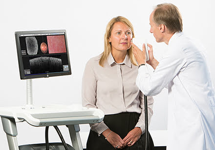
Jon Holmes, co-founder and CTO of Michelson Diagnostics Ltd., uses a clinical OCT scanner for dermatologic procedures. Courtesy of Michelson Diagnostics Ltd.
Nishant Mohan, vice president of the OCT division at Wasatch Photonics in Durham, N.C. There he leads development and commercialization of optical coherence tomography products. He is a former research fellow and senior R&D engineer for optical and medical entities. He studied electronics and communication engineering at the Indian Institute of Technology in Guwahati, India, and holds a Ph.D. in biomedical engineering from Boston University. He also serves on SPIE’s committee for public policy.
Dr. Richard B. Rosen, who serves at the New York Eye and Ear Infirmary of Mount Sinai as vice chair of the ophthalmology department, director of ophthalmology research, surgeon director of ophthalmology, and chief of retinal services. He is also a professor of ophthalmology at the Icahn School of Medicine at Mount Sinai and a visiting professor of applied optics in the School of Physical Sciences at the University of Kent (Canterbury, England). He obtained his medical degree from the University of Miami School of Medicine.
Igor Zhurminsky, a project leader at the Swiss Center for Electronics and Microtechnics (CSEM) SA Thin Film Optics Muttenz Center. He specializes in fabrication and application of micro and nanotechnologies, material sciences, industrial holography, holographic interferometry, diffraction optical elements, and laser technology. He holds a Ph.D. in physics from Moldova State University.
Are there new applications emerging for OCT technology? What are they?
Holmes: In dermatology, an emerging application is presurgical mapping of tumor margins, especially for Mohs surgery [a microscopically controlled surgical procedure that is used to treat skin cancers] for nonmelanoma skin cancer. The idea is to use OCT to image and demarcate the extent of a cancer prior to it being cut out, instead of the current approach of simply to guess the borders and then very often have to re-operate when a part of a tumor is left behind. OCT tumor mapping could make the process much more efficient and therefore more cost-effective, and the rising tide of skin cancer resulting from a sun-worshipping lifestyle and aging population will drive growth of this application.
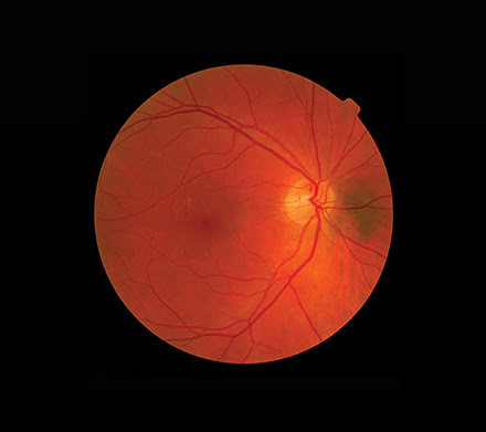
Optical coherence tomography (OCT) is most often used in ophthalmology for diagnosis and monitoring of retinal disease. But now this technology is expanding into new applications such as dermatology.
Another exciting new application for OCT in dermatology is guiding aesthetic treatments (especially laser treatments), such as removing unsightly blood vessels and wrinkles, and performing skin rejuvenation treatments. At the moment, most such treatments rely on the user’s experience for best results. There is often a trade-off between better results from aggressive treatment and safety for the patient. But we have some users who use OCT to image the skin of their patients and then set their lasers according to what they see. They believe that they are getting better results with a good safety margin. I expect this area to grow rapidly.
Mohan: OCT has been primarily used in ophthalmology for diagnosis and monitoring of retinal disease. There are several new applications for the technology that are coming into their own. Several medical technologies such as x-ray and ultrasound have found their way into commercial material inspection. OCT becoming an indispensable tool for certain nondestructive testing applications is not a matter of “if,” but “when.” Success of OCT in diagnostic ophthalmology is likely to have spill-over effects on other segments of ophthalmology, including different types of surgical procedures and neural disease where eyes can act as a window to the brain.
Rosen: OCT technology has the potential to provide functional and metabolic information, as well as cellular–level structural anatomy. The rapid development of OCT angiography is a prime example of functional information derived from OCT. As the speed of acquisition and processing OCT-A improves, the promise of variable speed imaging as pioneered by James Fujimoto, a professor in the Biomedical Optical Imaging and Biophotonics Group at MIT, and Dr. Nadia Waheed and Dr. Jay Duker, both from the New England Eye Center, may make the technique increasingly quantitative for characterizing blood flow.
Visible-wavelength OCT being developed by Hao Zhang, Ph.D., an associate professor of biomedical engineering at Northwestern University, and colleagues is currently being tested for its ability to provide metabolic information in the form of subtraction oximetry. This will provide clinicians with a bio-energetic picture of the components of a tissue, whose change in status typically precedes structural change.
Adaptive-optics OCT has been in development for years, but is only recently yielding groundbreaking new views of retinal ganglion cells in the inner retina and details of the choriocapillaries of the inner choroid, in the laboratories of Don Miller, Ph.D., a professor in Indiana University’s School of Optometry, and Robert Zawadzki, Ph.D., an assistant research professor in the department of ophthalmology and vision science at the University of California, Davis. These previously unavailable cellular views will further our understanding of the development and the progression of chronic diseases such as glaucoma and macular degeneration.
Expanded-field OCT imaging will also change clinical management. Higher speed systems — such as swept-source and faster spectrometers — invite faster data collection and the opportunity for wider acquisition at reasonable imaging times. Clinicians such as Dr. Venera A. Shaimova, a senior researcher at the Chelyabinsk State Laser Surgery Institute, and Dr. Netan Choudry, a clinical teacher in the University of Toronto’s department of ophthalmology and vision sciences, have recently demonstrated independently the ability to capture lesions in the peripheral retina, which will improve our ability to identify and treat disease in these areas more precisely and successfully.
Zhurminsky: [Some emerging technologies CSEM is seeing include] security applications, measurements of microlenses, and measurements of encapsulation thickness.
What are the biggest challenges for new advances in OCT?
Holmes: This depends on what the new advance is specifically targeting. If it is aimed at an existing OCT application, but with smaller/lower-cost technology, then the challenge is not to compromise on performance. Clinicians will not adopt a new device if the image quality is noticeably worse than what they have been using for years and has been proven in large expensive trials. For example, I would like to see lower-cost swept-source lasers with the same specifications as today’s state-of-the-art.
But if the new advance in OCT is targeting a new clinical application, then one must always consider whether it is affordable and can be paid for — the cost per patient versus existing gold standard practice, the number of patients per site per annum, and whether there is a route to reimbursement for the scan procedure. Finally, any new technology has to be proven in large clinical trials. These challenges are very tough!
Mohan: One has to consider both research and commercial aspects in this case. Academic research in OCT is largely driven by public funding; downward pressure on such funding can have a negative impact on research advancements. Commercially, OCT will face challenges to maintain continued growth in the existing markets. Pressure to reduce medical costs will likely result in lower reimbursement rates and reduced adaption of the technologies like OCT. OCT developers will need to show continuous improvement in health outcomes to sustain the market growth.
Rosen: Among the biggest challenges for new advances in OCT is the economic inertia encountered by manufacturers and the end users. The cost of development and the resistance of the market to pay for new imaging are major hurdles. In addition to providing new information, developers must demonstrate major improvements in decision-making and outcomes. The lack of availability of therapeutics, which can take advantage of the information revealed by the new imaging modality, can delay acceptance for extended periods of time.
Zhurminsky: It would be nice to enlarge application fields to sub-micron range by increasing resolution in XYZ to nanometer scale. At the moment, XY resolution is 1.5625 µm, and Z is 0.4 µm. Measurements should be adapted to the sample — for example, a sample with a high reflection or high diffusion surface. This adaptation needs some special tricks such as a deposition of nanometer thickness of materials, which decreases a reflection or some modification in the measuring options in the proper device. It would probably be more efficient to fulfill this adaptation by software tools.