
Tissue Scaffolding Made from Laser-Patterned Silk
Hydrogels made of silk protein have emerged as a promising platform for tissue engineering thanks to their transparency, which allows laser patterning deep below the material's surface.
Silk protein is a soft biomaterial that supports cell growth and allows cells to penetrate deep within it. Researchers at Tufts University have demonstrated that femtosecond laser pulses with energies <2 nJ can be used to create 3D structures as small as 10 μm that facilitate the flow of oxygen and nutrients through the material.
"Because the femtosecond laser pulses allow us to target specific regions without any damage to the immediate surroundings, we can imagine using such micropatterning to controllably design around living cells, guide cell growth and create an artificial vasculature within an already densely seeded silk hydrogel," said professor Fiorenzo G. Omenetto.
Existing patterning techniques allow for the production of random, microscale pores and the creation of channels that are hundreds of microns in diameter, with few options in between, the researchers said.
Unlike most 3D printing, the new technique does not require photoinitiators, or compounds that promote photoreactivity but are typically bio-incompatible. Instead the structures are created through multiphoton absorption.
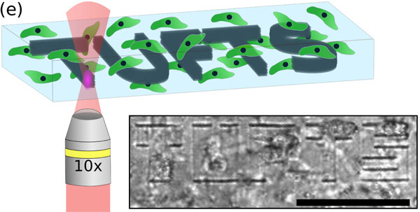
Illustration of laser-based micropatterning of silk hydrogels. The transparent gels enable laser photons to be absorbed more than 10 times deeper than in other materials, without damaging the cells surrounding the "Tufts" pattern. Courtesy of M.B. Applegate/Tufts University.
The hydrogel's transparency allows laser photons to penetrate up to 1 cm below the surface — more than 10 times deeper than other materials.
Tested in vitro and in a preliminary in vivo study in mice, the laser treatment can be done while keeping the cell culture sealed and sterile.
Funding came from the U.S. Office of Naval Research. The research was published in PNAS (doi: 10.1073/pnas.1509405112).
Published: September 2015