Anesthetics used in prolonged medically induced comas may trigger cognitive deficits. Examining effects at the neuronal level may help doctors to chart a safer course.
MICHAEL WENZEL, BONN UNIVERSITY HOSPITAL, AND RAFAEL YUSTE, COLUMBIA UNIVERSITY
The formation of synapses in the circuitry of the brain, and especially the solidification and disappearance of synaptic structures, mediates how people learn, memorize, and forget. Since the introduction of two-photon imaging to neuroscience in the 1990s, it has been used to image synaptic connections in the brains of living animals. Two-photon imaging has also revealed changes in neural functioning that underlie neuropsychiatric diseases such as epilepsy, multiple sclerosis, and schizophrenia. A recent study of two-photon synaptic imaging of mouse brain during prolonged anesthesia revealed major structural changes in synapses1, highlighting the utility of two-photon microscopy to provide data that could justify shifts in patient care, specifically in intensive care medicine.
A focus on the ICU
As a critical life-saving measure, prolonged medically induced coma (pMIC) is carried out millions of times annually on patients in intensive care units (ICUs) around the world. Throughout the
COVID-19 pandemic, intensive care has often been the epicenter of the public health crisis as the number of severely affected SARS-CoV-2 patients who required mechanical respiratory support outgrew the capacity of ICUs in many parts of the world. The long-term health effects on patients who survived severe COVID-19 and who required mechanical respiratory support may not be understood for many years.
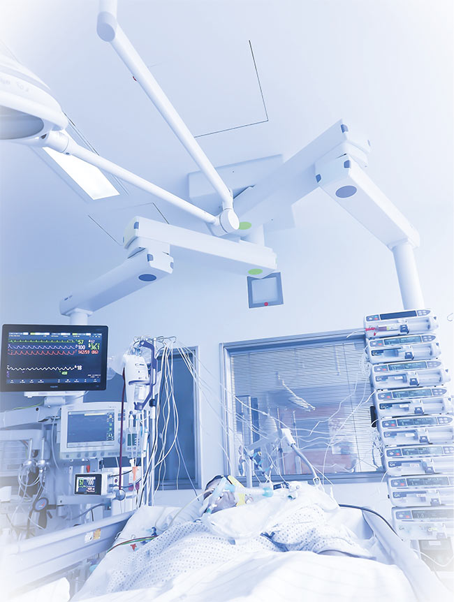
A critically ill patient in a prolonged medically induced coma. Such comas are stably maintained using a combination of anesthetics, including the inhalatory agent isoflurane, the use of which is experiencing a renaissance in hospital intensive care units. Courtesy of Michael Wenzel/Bonn University.
The renewed attention on intensive medical care has put a spotlight on some common ICU procedures, such as the
use of a combination of anesthetic agents to maintain medically induced coma. These protocols have been built upon decades of clinical experience, but
often without clear scientific validation.
Up to one-third of ICU patients requiring mechanical respiratory support develop cognitive impairment2. It has been speculated that this development may be attributable not only to the respective underlying diseases, but also to the direct effects of anesthesia itself. While there is evidence that anesthetics can lead to synaptic changes and cognitive impairment in children — who adapt rapidly to changing environmental inputs — until recently, there was no evidence of such effects in adults. Further, despite the assumption in the medical community that the effects of pMIC on the brain become more pronounced the longer the procedure lasts, the longest prolonged medically induced coma that was studied previously was just 6 h. In intensive medical care, however, medically induced comas are routinely maintained for days and sometimes weeks or months.
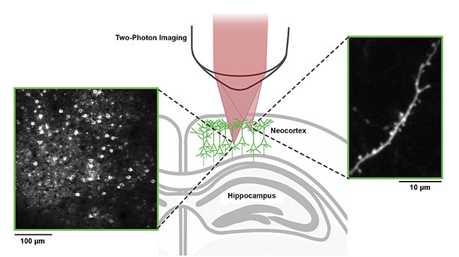
Two-photon imaging enables the mapping of neurons and synapses in living brain. A fluorescently labeled synapse-bearing dendritic compartment (right box), located approximately 25 µm below the mouse brain surface. A typical field of view spanning several hundred micrometers (left box). The imaging focal plane is located approximately 150 µm below the brain surface, enabling hundreds of individual neuronal cell bodies to be readily identified and their activity monitored over time. Courtesy of Michael Wenzel/Bonn University.
Because the reasons for cognitive impairment in critically ill patients after pMIC have remained incompletely understood, systematic therapeutic strategies for minimizing disruptions to brain circuitry during extended anesthesia have not been developed, and patient-tailored clinical guidelines with regard to anesthetic combinations do not currently exist. This lack of knowledge has immense health-related and socioeconomic consequences for patients who survive a critical medical condition involving pMIC, as well as ramifications for patients’ families, medical caregivers, the health care system, and society as a whole.
Technical challenges
The reasons for this lack of knowledge include the difficulty of differentiating the direct effects of pMIC from the effects of medical conditions that precipitated the procedure, along with the difficulty of studying brain changes at synaptic resolution in humans. The recent development of radioactively labeled compounds that target synapses has made it possible to make indirect assumptions of brain-wide synaptic density in the human brain using positron emission tomography3. But this technique suffers from poor spatial resolution, hindering fine-scale investigations of synaptic changes.
Since the advent of two-photon fluorescence microscopy, it has become technically possible to study neural circuitry within the living brain of animals at subcellular resolution and over a prolonged period of time. While current microscopy imaging approaches still suffer from limited depth resolution (at best, up to few millimeters beneath the brain surface), strong progress is being made.

Chronic in vivo two-photon imaging enables the monitoring of synaptic dynamics through chronically implanted cranial windows. The dense dendritic
arborization of layer 5 pyramidal neurons in vivo, ~10 to 20 µm below the mouse brain surface (green box). A magnified inset shows an individual dendrite containing dozens of dendritic spines within only dozens of micrometers (red box). A more-magnified inset shows high-fidelity tracking of even individual
synaptic dynamics within the alive brain (blue box). The hollow arrows mark where individual synapses have disappeared, while the filled arrow shows
a new synapse. Illustration courtesy of Michael Wenzel. Data courtesy of Michael Wenzel/Yuste Laboratory/Columbia University.
Despite such technical advancements, synapse-level studies of the effects of pMIC on the intact brain have lagged. Neuroscientists have had little motivation to study the procedure in mice, partly because early studies researching synapse dynamics across developmental stages in mice provided evidence for increasingly stabilized synaptic connections in adulthood. In nondiseased adult mouse cortex, the overwhelming majority (>95%) of dendritic spines (a structural correlate of neuronal synapses) remained stable across a one-month interval, with a half-life greater than 13 months4. When cortical synapses were monitored across a 6-h period of pMIC in adult mice in a recent study, no significant changes in dendritic spine dynamics were found5. Maintaining mice in a medically induced coma under stable conditions for prolonged periods is also difficult. In comparison to human patients, mice have a much-accelerated metabolism, heart rate, and breathing rate, along with a ratio of body weight to surface area that predisposes them to rapidly lose body temperature during pMIC-induced resting conditions.
From bedside to bench
Answering questions about the direct effects of pMIC on the brain remained a pressing goal in the medical community because of the long periods of time in which humans are often routinely placed in medically induced coma. An experimental framework was recently established that enables stably maintained pMIC in mice (for up to 40 h) in combination with chronic synaptic-resolution two-photon imaging of cortical synapses and repeated behavioral assessment1.
It was found that, contrary to prevailing thought, pMIC has significant side effects on the synaptic architecture of the brain, regardless of the age of the animal. During pMIC, the brain forms significantly more synapses than it does under normal conditions. After discontinuation of the procedure, the brain disproportionally loses synaptic connections. Importantly, half of the synapses formed during the procedure remained stable for several days afterward, suggesting that these synapses may indeed become functional. Since no specific learning experience takes place during a prolonged medically induced coma, this unspecific, abnormal formation of neural connections may contribute to cognitive impairment in pMIC patients because the additional synapses could interfere with normal neural functioning.
From bench to bedside
What are the real-world implications of these findings? The pilot two-photon imaging study identified significant structural and functional side effects of pMIC, showing that the synaptic architecture of the adult brain is far less stable than previously thought. The study exclusively used isoflurane, an inhalatory anesthetic, due to its fast kinetics that enable rapid adjustability of anesthetic depth. The findings make the case for a systematic investigation of other synapse-level effects and related cognitive deficits triggered by common treatments in intensive care, including propofol and benzodiazepines, as well as particular combinations of anesthetics.
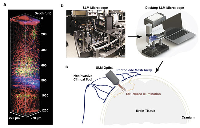
3D volume reconstruction of live mouse brain using three-photon imaging (a). A portable holographic microscope for potential multiphoton imaging in humans. A typical two-photon microscopy setup in a lab (b, left). An optically equivalent microscope that uses a spatial light modulator (SLM) to replace most of the optical elements (b, right). Depiction of an SLM-based microscope mounted on the skull for deep brain imaging (c). Image (a) reprinted with permission from Reference 10/CC BY-NC 4.0; (b) and (c) adapted with permission from Reference 6.
It is possible that particular anesthetics or combinations thereof may have less detrimental effects on brain synaptic architecture than others. The findings herein may encourage more research on this subject, as well as research into adjuvant therapies that may help to maintain brain structure during pMIC. Patients in medically induced comas receive physiotherapy to maintain the mobility and function of their joints and muscles, yet no adjunctive cognitive or brain-activity therapy exists for the anesthetized brain.
Outlook and challenges
A better understanding of the effects of prolonged medically induced coma on brain synapses could allow for patient-tailored anesthetic regimens and boost research on adjuvant therapeutic strategies to improve the cognitive retention of pMIC patients. In vivo imaging with two-photon microscopy could be an optimal tool in advancing toward this goal. Ideally, pMIC-associated changes in brain synaptic architecture would be studied directly in human patients.
On a technical level, laser-scanning optical techniques such as two-photon imaging could be used in humans without causing harm because the method uses pulsed laser light in the near-infrared range (750 to 2500 nm), which allows penetration and recording of living tissue without damaging it6. Spatial light modulators (SLMs) could be used to miniaturize imaging technology, which could be mounted on the skull for deep brain imaging7.
However, several factors make it difficult to perform in vivo two-photon imaging in humans. First, the integration of two-photon microscopy into the clinical workflow greatly extends procedure duration due to additional imaging, which could affect patient well-being. Second, the technique offers limited depth resolution. The identification and characterization of nanocrystals that offer suitable properties for higher-order nonlinear light absorption (such as five-photon absorption) could be applied in biological imaging to increase depth penetration and maintain synaptic spatial resolution. However, such approaches are still in the earliest stages of development8. Third, to reduce the risk of harming the sampled tissue, more research on nontoxic fluorescent indicators for labeling neurons in the human brain is necessary (e.g., using NIR-fluorescent dye-doped nanoparticles, BODIPY, or coumarin derivatives)9.
Despite these challenges, multiphoton imaging technology could advance knowledge of brain synapse dynamics and drive clinical-level innovations in intensive care medicine. Therapeutic interventions based on data provided by this technology could help to improve clinical outcomes for patients who receive prolonged general anesthesia.
Meet the authors
Michael Wenzel, M.D., Ph.D., is a research clinician specializing in neurology, and junior group leader in the Department of Epileptology at Bonn University Hospital. He obtained his medical degree and an experimental doctoral degree from Ludwig-Maximilians-Universität in Munich. Between 2014 and 2018, he worked as a postdoctoral fellow in Rafael Yuste’s laboratory at Columbia University. He is a fellow of the Hertie Network of Excellence in Clinical Neuroscience; email: [email protected].
Rafael Yuste, M.D., Ph.D., is professor of biological sciences at Columbia University. He obtained his medical degree from Universidad Autónoma de Madrid. After working in Sydney Brenner’s laboratory in Cambridge, England, he worked as a doctoral student with Menachem Katz in Torsten Wiesel’s laboratory at Rockefeller University, and then worked as a postdoctoral student with David Tank at Bell Laboratories. He joined Columbia University in 1996 and is currently director of its Neurotechnology Center. Yuste led a group of researchers who proposed the U.S. BRAIN Initiative, and he helped coordinate the International BRAIN Initiative. He is involved in ethical guidelines for neurotechnologies (“NeuroRights”); email: [email protected].
Acknowledgments
This work was supported by NEI (R01EY011787), NINDS (R01NS110422; R34NS116740), NIMH (R01MH115900), BONFOR (2019-2-04), and the Hertie Foundation (P1200008).
References
1. M. Wenzel et al. (2021). Prolonged
anesthesia alters brain synaptic architecture. Proc Natl Acad Sci, Vol. 118,
No. 7, e2023676118, www.doi.org/10.1073/pnas.2023676118.
2. P.P. Pandharipande et al. (2013). Long-term cognitive impairment after critical illness. N Engl J Med, Vol. 369, No. 14,
pp. 1306-1316, www.nejm.org/doi/abs/10.1056/nejmoa1301372.
3. S.J. Finnema et al. (2016). Imaging synaptic density in the living human brain. Sci Transl Med, Vol. 8, No. 348, pp. 1-10,
www.doi.org/10.1126/scitranslmed.aaf6667.
4. J. Grutzendler et al. (2002). Long-term dendritic spine stability in the adult cortex. Nature, Vol. 420, No. 6917, pp. 812-816, www.doi.org/10.1038/nature01276.
5. G. Yang et al. (2011). Transient effects of anesthetics on dendritic spines and filopodia in the living mouse cortex. Anesthesiology, Vol. 115, No. 4, pp. 718-726, www.doi.org/10.1097/aln.0b013e318229a660.
6. W. Chen et al. (2017). Giant five-photon absorption from multidimensional core-shell halide perovskite colloidal nanocrystals. Nat Commun, Vol. 8, Article No. 15198, www.doi.org/10.1038/ncomms15198.
7. V. Nikolenko et al. (2010). A portable laser photostimulation and imaging microscope. J Neural Eng, Vol. 7, No. 4, www.doi.org/10.1088/1741-2560/7/4/045001.
8. W. Yang and R. Yuste (2017). In vivo imaging of neural activity. Nat Methods, Vol. 14, No. 4, pp. 349-359, www.doi.org/10.1038/nmeth.4230.
9. M. Bojtár et al. (2019). Green-light activatable, water-soluble red-shifted coumarin photocages. Org Lett, Vol. 21, No. 23, pp. 9410-9414, www.doi.org/10.1021/acs.orglett.9b03624.
10. Y. Hontani et al. (2021). Multicolor three-photon fluorescence imaging with single-wavelength excitation deep in mouse brain. Sci Adv, Vol. 7, Issue 12, www.doi.org/10.1126/sciadv.abf3531.