A novel superresolution nanoscope allows 3D imaging of an entire mammalian cell and its cellular constituents at a resolution that is 20 to 50 times higher than conventional microscopy, with imaging depth improved approximately tenfold over state-of-the-art iPALM and 4Pi-SMSN implementations. Until now, resolving details at this level was only possible using electron microscopy, which requires samples to be treated.
The whole-cell 4Pi single-molecule switching nanoscope — called W-4PiSMSN and developed by a team from Purdue University, Yale University, the University of Cambridge, the Jackson Laboratory, Howard Hughes Medical Institute and the University of Oxford — allows volumetric reconstruction with 10- to 20-nm isotropic resolution of approximately 10-μm-thick samples. The novel nanoscope permits ultrahigh-resolution 3D imaging of virtually any subcellular structure, allowing researchers to resolve details smaller than the wavelength of light.
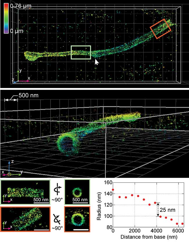
Using a new 3D superresolution instrument, researchers captured this visualization of a "primary cilium," the antenna of the cell. Courtesy of Huang et al./Cell.
The W-4PiSMSN incorporates deformable mirrors using two objectives, one above and one below the sample, and uses a set of algorithms to pinpoint molecular positions of proteins deep inside cells, resolving features deep below the surface of the sample.
Researchers have demonstrated the wide applicability of W-4PiSMSN by imaging complex molecular architectures ranging from bacteriophages (viruses about 50 nm in diameter) to nuclear pores, cilia and synaptonemal complexes in large 3D cellular volumes. The development of W-4PiSMSN extends the application range of 4Pi-based SMSN to the imaging of organelles that span large volumes, exemplified by the mitochondrial network, the nuclear envelope, and synaptonemal complexes, which researchers were able to capture in virtual entirety.

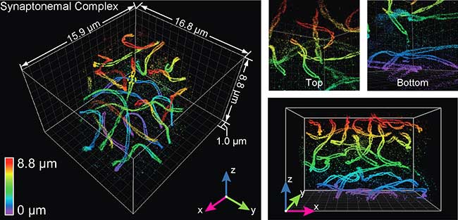
The technology was used to visualize a mouse spermatocyte, revealing with unprecedented clarity the "twisting paired lateral elements" of synaptonemal complexes, which link chromosomes together. Courtesy of Huang et al./Cell.
The advance could reveal biological phenomena never before seen, bringing novel medical insights.
"One goal is to further push the envelope in the direction of live-cell and tissue imaging, two major roadblocks of modern superresolution techniques, and therefore allow visualization of cellular functions live in their physiological conditions at the nanoscale,” said Fang Huang, professor of biomedical engineering at Purdue.
"We are interested in using our developments to study the cytokinetic apparatus, a core machinery during cell division," he added.
W-4PiSMSN is a versatile and powerful tool that promises a new perspective on how proteins distribute across entire organelles throughout whole cells, an as yet unmet challenge in cell biology.
The research was published in Cell (doi: 10.1016/j.cell.2016.06.016).