|
Wednesday, November 27, 2019
|
|

|
|
|
Monthly newsletter focusing on how light-based technologies are being used in the life sciences. Includes news, features and product developments in lasers, imaging, optics, spectroscopy, microscopy, lighting and more. Manage your Photonics Media membership at Photonics.com/subscribe.
|
|
|
|
sponsor
|

|
|
The Eyes Behind Surgical Robots
The first robotic surgery took place in 1985 when the PUMA 560 was used in a stereotaxic operation in which computed tomography (or x-ray) was used intraoperatively to guide a robot as it inserted a needle into the brain for biopsy. In the late 1980s and early 1990s, robotic systems began to be used for laparoscopic surgery, in which a flexible optical instrument was inserted into the body and used to guide surgeons through hard-to-reach areas, from the pelvis to the chest cavity. Fast forward two decades, and today’s surgical robots are more precise and user-friendly, and capable of highly delicate microsurgeries, thanks to advancements in optical technology that have allowed for safe and effective intraoperative imaging. From a patient’s perspective, one of the biggest benefits of robot-assisted minimally invasive surgery is the reduced trauma, which enables quicker recovery times when compared to open surgery.
|
|
|
|
|
|
Deconvolution Helps Break Down Imaging Barriers
Deconvolution is a computationally intensive image processing technique used to improve the contrast and sharpness of images captured with a light microscope. Light microscopes are diffraction limited, which means they are unable to resolve individual structures unless they are more than half the wavelength of light away from one another. Each point source below this diffraction limit is blurred by the microscope into what is known as a point spread function (PSF). With traditional wide-field fluorescence microscopy, out-of-focus light from areas above or below the focal plane causes additional blurring in the captured image. This becomes a problem when trying to identify or measure structures inside thicker samples.
|
|
|
|
|
|
Compressive Raman Bioimaging: The Need for Speed in Medicine
It has been widely demonstrated that Raman spectroscopy provides powerful information for biomedical applications1, including real-time tumor surgery imaging, breast cancer diagnostics, detection of Alzheimer’s plaques, and histopathology. These applications detect and quantify various biomolecules through vibrational spectroscopy analysis, such as proteins, lipids, and nucleic acids. All of these examples present possibilities for using Raman-based imaging, with the majority of proof-of-principle experiments being performed ex vivo or with the sample spatially frozen. However, in biology, nothing is really fixed in space; because of inherent organelle movement, or just a simple heartbeat, bioimaging often needs to be faster than any movement artifacts. So speeding up Raman imaging is a major challenge tackled by numerous groups worldwide.
|
|
|
|
|
|
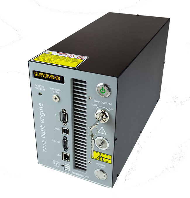 ZIVA Light Engine®
Lumencor Inc.
The ZIVA light engine is optimized for coupling into narrow bore fibers (≤100 µm) providing a system with ultra high radiance from their small emitting area, much brighter than its predecessor the CELESTA light engine.
Visit Website
Request Info
|
|
|
 OEM LED Illumination Service
CoolLED Ltd.
CoolLED is expanding its OEM LED Illumination business to give developers of high-content screening and slide scanning systems a competitive edge in the market. Since introducing the first commercially available LED illumination system in 2006, the company has led the way in light source technology for fluorescence...
Visit Website
Request Info
|
|
|
|
|
|
 Optical Biomedical Imaging
Photonics Media
At last, a reference work has been compiled that offers in one place a broad survey of technologies, applications and markets for optical biomedical imaging, as only Photonics Media could produce it. This collection is a practical resource for those engaged in the research and development of relevant technologies.
Visit Website
Request Info
|
|
|
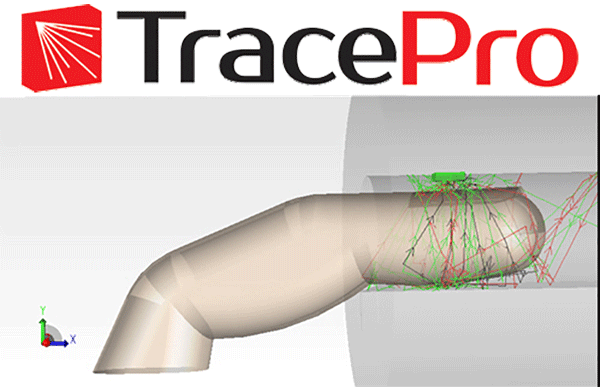 TracePro Enhances Life Science Research
Lambda Research Corp.
Life Science Research & Discovery Optical modeling for life science research requires an extraordinary degree of interdisciplinary collaboration among medical doctors, life scientists, and biomedical design engineers.
TracePro offers:
- Intuitive Modeling Environment
- Collaboration-Enhancing
Visit Website
Request Info
|
|
|
|
|
|
 Compact and High-Power LED Illumination
Excelitas Technologies Corp.
The X-Cite mini+™ is the new go-to white-light LED light source for fluorescence imaging applications. High-power performance, compact size, and economically priced, the X-Cite mini+ is the perfect choice for routine fluorescence imaging with enough power to replace mercury sources in budget-constrained labs and clinical facilities.
Visit Website
Request Info
|
|
|
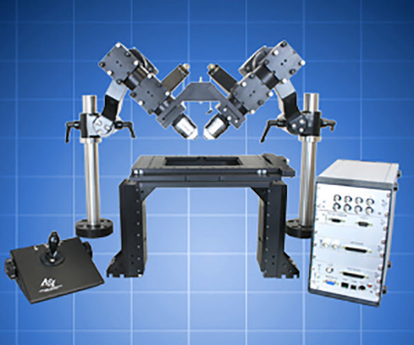 Dual Light Sheet Microscopy
Applied Scientific Instrumentation Inc.
ASI’s Dual Selective Plane Illumination Microscopy for Cleared Tissue (ct-dSPIM) is one of many light sheet microscope configurations possible using our modular components. This flexible and easy-to-use Selective Plane Illumination Microscopy (SPIM) implementation allows for dual views of large samples such as cleared tissue (ct).
Visit Website
Request Info
|
|
|
|
|
|
AI Improves Quality and Usability of Optoacoustic Imaging
To improve image quality in low-cost optoacoustic devices with only a small number of ultrasonic sensors, researchers at ETH Zurich and the University of Zurich developed a framework for the efficient recovery of image quality from sparse optoacoustic data using a deep convolutional neural network and demonstrated their approach with whole body mouse imaging in vivo.
|
|
|
|
|
|
A new, noninvasive optical imaging system promises to improve diagnosis and treatment for dry eye disease. Called the Tear Film Imager, the device has the ability to perform spectral measurements across a large field of view in a few seconds and can acquire fast and consistent measurements from a blinking human eye. The Tear Film Imager uses an eye-safe halogen light to illuminate the eye and then analyzes the full spectrum of light reflection over time and space. These spectral measurements are used to reconstruct the structures found in the front of the eye, allowing accurate measurement of the tear film inner layers, especially the aqueous sublayer, which plays an important role in dry eye but has been difficult to analyze with other methods.
|
|
|
|
Engineers at Duke University used machine learning to develop a microscope capable of adapting its lighting angles, colors, and patterns while teaching itself the optimal settings needed to complete a diagnostic task. In a proof-of-concept study, the microscope simultaneously developed a lighting pattern and classification system that allowed it to quickly identify red blood cells infected by the malaria parasite. According to the research team, it performed these tasks more accurately than trained physicians and other machine learning approaches.
|
|
|
|
Vision Systems for Deep Learning
Thu, Dec 12, 2019 1:00 PM - 2:00 PM EST
Algorithms for artificial intelligence are improving rapidly. In the medical and life sciences fields, in particular, many classification problems that were once considered to be “nonsolvable” by machines can now be solved with an impressive level of accuracy and robustness. This webinar, presented by Basler, will give you an overview of three different types of vision systems that can be used to deploy a trained neural network in the medical and life sciences fields: Embedded; PC-based; and FPGA-based. The webinar will discuss how these system architectures differ in their total cost of ownership, in the engineering effort required to deploy them, and in overall system performance.
|
|
|
|
Features
STED Nanoscopy for Cell Biology, Spectroscopy and the Environment, Lasers in Microscopy
Photonics Media is currently seeking technical feature articles on a variety of topics for publication in our magazine BioPhotonics. Please submit an informal 100-word abstract to Senior Editor Doug Farmer at [email protected] or use our online submission form www.photonics.com/submitfeature.aspx.
|
|
|
|