
Cooling Stage
Linkam Scientific Instruments Ltd.Request Info
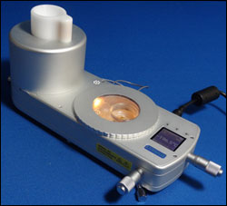
Linkam Scientific Instruments has developed a stage for cryo-correlative light electron microscopy (cryo-CLEM) used in imaging of cells at cryo temperatures. It was designed in collaboration with Leiden University Medical Centre.
Cryo-CLEM is the correlation of images captured with a cryo stage on a fluorescent light microscope and images of the same sample observed with transmission electron microscopy (TEM).
 The correlative stage is being used as part of a work flow that starts with cells expressing cancer-related genes and ends with imaging in synchrotrons located in Oxford, Berlin and Barcelona, Spain. Within the synchrotrons, the scientists can image the cells using soft x-ray tomography.
The correlative stage can hold samples at a stable −196 °C, enabling scientists to study TEM grid samples at 100× magnification, identifying areas of further interest and facilitating the movement of the analyzed grids to the TEM. With automated liquid nitrogen control, heated optics and a digital display, the unit is a compact and efficient system.
It is used to study cells that are undergoing autophagy, a process involving degradation and recycling of unnecessary or dysfunctional cell components. It also is being used in a process for imaging fluorescent proteins in cells as close to their living state as possible, considering that they must be placed in a vacuum to take high-magnification images. The cells are grown on thin carbon films attached to a 3-mm-diameter gold grid and frozen in liquid ethane at −174 °C. The correlative stage is used to image the fluorescence in the cells while they are still frozen. The frozen grids are then shipped to the synchrotron, where they are put into the soft x-ray microscope so the structure of the entire cell can be imaged in 3-D.
http://www.linkam.co.uk
/Buyers_Guide/Linkam_Scientific_Instruments_Ltd/c18051
Published: December 2012
REQUEST INFO ABOUT THIS PRODUCT
* First Name:
* Last Name:
* Email Address:
* Company:
* Country:
Message:
When you click "Send Request", we will record and send your personal contact information to Linkam Scientific Instruments Ltd. by email so they may respond directly. You also agree that Photonics Media may contact you with information related to this inquiry, and that you have read and accept our
Privacy Policy and
Terms and Conditions of Use.
Register or login to auto-populate this form:
Login
Register
* Required