
Diffusion Measurement Module
Olympus Europa SE & Co. KGRequest Info
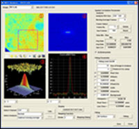
Olympus Life Science Europa GmbH has introduced a diffusion measurement module for its ASW 2.1 software, designed specifically for use with its FluoView FV1000 confocal microscope system for live-cell imaging.
 The software package provides users with the flexibility to perform three types of diffusion study: point fluorescence correlation spectroscopy, raster scan image correlation spectroscopy (RICS) and fluorescence recovery after photobleaching (FRAP). It is suitable for use in a variety of cell biology and biophysics applications.
Intracellular diffusion is frequently analyzed to assess the movements, interactions and microenvironments of intracellular molecules to determine the patterns and rates of molecular movement.
In point fluorescence correlation spectroscopy, data is obtained from a single location using point scanning, enabling recording of fluctuations in fluorescence intensity. The number of moving particles within the confocal volume is therefore easily calculated. With its high temporal and spatial resolution, this technique provides data on the diffusion constant or number of molecules, and enables analysis of cross correlations between different molecules.
For RICS, the software provides diffusion measurements in various cellular regions and presents the user with a spatial mapping of diffusion time constants. As a result, users can easily define small regions of interest and subsequently create a diffusion map from a series of 2-D images. A wide range of intracellular structures, from molecules in solution to cell membrane proteins, can therefore be accurately measured. Resulting data can be expressed as either a diffusion constant or as the number of available molecules.
Researchers commonly use FRAP to analyze the diffusion constant in a particular region of the cell, as molecules diffuse into the area after photobleaching.
The diffusion measurement package enables users of the Olympus FluoView FV1000 to easily control and measure the diffusion constant.
http://www.olympus-ims.com
/Buyers_Guide/Olympus_Europa_SE_Co_KG/c10690
Published: August 2010
REQUEST INFO ABOUT THIS PRODUCT
* First Name:
* Last Name:
* Email Address:
* Company:
* Country:
Message:
When you click "Send Request", we will record and send your personal contact information to Olympus Europa SE & Co. KG by email so they may respond directly. You also agree that Photonics Media may contact you with information related to this inquiry, and that you have read and accept our
Privacy Policy and
Terms and Conditions of Use.
Register or login to auto-populate this form:
Login
Register
* Required