
Life Sciences Software
Olympus Europa SE & Co. KGRequest Info
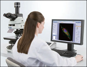
Olympus Europa Holding GmbH has announced version 1.7 of its cellSens life sciences software featuring the “well navigator” solution for multiwell imaging experiments.
 It offers high-dynamic-range imaging, 2-D deconvolution and a new database for streamlining management of large data repositories with many users. The software supports additional models of high-sensitivity cameras and is compatible with the company’s real-time controller for performing total internal reflection fluorescence.
The software blends the company’s frames, cameras and optics to form a complete microscopy system. When the user is working with challenging samples containing both very light and dark staining, the high-dynamic-range mode can bring out all the details with one click. The 2-D deconvolution tool improves images by quickly applying a true deconvolution on a single image, without the need for acquiring a Z-stack, for clear images of samples stained with multiple fluorochromes.
The software includes customizable pre-sets for a variety of plate formats and panoramic imaging of each well. It works with the proprietary ZDC focus-compensation unit, which can automatically maintain perfect focus without user intervention.
When studying fast dynamic processes, acquisition accuracy is a must for reliable quantitative analysis. The software supports a dedicated real-time processor that facilitates advanced imaging via microsecond synchronization of the camera, light source and filter units. The database has been redesigned to provide centralized data storage management, optimized for multi-user access. Large groups or different institutions can collaborate by securely sharing research data, while maintaining quick and easy access through customizable databases.
User interface enhancements include the ability to use existing images to set the microscope system to the same acquisition settings, ensuring consistent analysis across many different samples. The new “dark skin” mode reduces monitor brightness when performing fluorescence imaging, while maintaining clear colored icons for intuitive ease of use.
For more information, visit: www.microscopy.olympus.eu
http://www.olympus-ims.com
/Buyers_Guide/Olympus_Europa_SE_Co_KG/c10690
Published: July 2012
REQUEST INFO ABOUT THIS PRODUCT
* First Name:
* Last Name:
* Email Address:
* Company:
* Country:
Message:
When you click "Send Request", we will record and send your personal contact information to Olympus Europa SE & Co. KG by email so they may respond directly. You also agree that Photonics Media may contact you with information related to this inquiry, and that you have read and accept our
Privacy Policy and
Terms and Conditions of Use.
Register or login to auto-populate this form:
Login
Register
* Required