
Light Sheet for Cleared Tissue
Applied Scientific Instrumentation Inc.Request Info
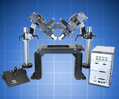 The ct-dSPIM is a flexible and easy-to-use light sheet microscopy configuration optimized for imaging large cleared tissue samples. The sample is mounted on a motorized XYZ stage and imaged via stage scanning. Two multi-immersion or other objective lenses are held in an upright “V” geometry for light sheet illumination and detection. Acquisition speed is usually camera-limited.
ASI manufactures the optomechanical elements, including the motorized stages and 2D galvos for creating and moving the light sheet. Lasers and sCMOS cameras are required to complete the system; users can procure these themselves, use the services of various system integrators selling ASI SPIM systems, or purchase them via ASI. The ct-dSPIM has been successfully used to image various cleared tissue samples including whole mouse brains and slices of cleared tissue.
ct-dSPIM Features
Image acquisition >10^8 voxels/sec
Sub-micron resolution in XYZ (sample-permitting)
Sample mounting in open dish
Image >5 mm deep into flat samples or up to 12 mm radius sphere
Media RI range from 1.33 to 1.56, aqueous or organic media
Modular and flexible setup
http://www.asiimaging.com/products/light-sheet-microscopy/dual-selective-plane-illumination-microscopy-for-cleared-tissue/
/Buyers_Guide/Applied_Scientific_Instrumentation_Inc/c1021
REQUEST INFO ABOUT THIS PRODUCT
* First Name:
* Last Name:
* Email Address:
* Company:
* Country:
Message:
When you click "Send Request", we will record and send your personal contact information to Applied Scientific Instrumentation Inc. by email so they may respond directly. You also agree that Photonics Media may contact you with information related to this inquiry, and that you have read and accept our
Privacy Policy and
Terms and Conditions of Use.
Register or login to auto-populate this form:
Login
Register
* Required