
QDI 302 Microscope Spectrophotometer
CRAIC TechnologiesRequest Info
SAN DIMAS, Calif., Feb. 2, 2010 – Organic light-emitting diodes (OLEDs) are being developed for the next generation of displays and light sources. The QDI 302 microscope spectrophotometer from Craic Technologies Inc. is designed to measure and compare the spectral output, intensity and color consistency of each of the microscopic pixels commonly found in OLED devices.
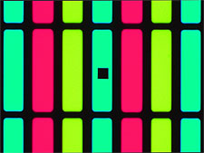 OLEDs have an emissive electroluminescent layer that consists of organic molecules in a supporting matrix. For displays, this layer is formed into millions of microscopic pixels in ordered rows and columns. As different organic compounds are used to generate different colors, pixels with various organic compounds can generate different colors for full color high-resolution displays. The advantage of OLEDs, as opposed to LCDs, is that the pixels combine both the light source and the color source, so that OLED displays are lighter and thinner and use less electricity. The QDI 302 spectrophotometer for microscopes measures the consistency of the intensity and the color of the optical emission across the device.
The spectrophotometer attaches to the open photoport of a microscope or probe station and enables the user to acquire images and spectra of microscopic sample areas quickly. Transmission, reflectance, polarization and fluorescence spectra of microscopic samples can be collected. When added to the appropriate microscope or probe station, the device can be used to measure the color and intensity of each pixel of an OLED display. Pixels can then be compared with one another for consistency, and maps of both the intensity and color can be generated for each device. The spectrophotometer can acquire spectra on the order of a few milliseconds, and entire OLED displays can be mapped quickly and accurately to ensure the consistency of both color and intensity across the entire device as well as from device to device.
Depending upon the microscope optics and sources, the spectral range is from the deep-UV to the near-infrared. High-quality spectra of submicron samples can be easily and nondestructively acquired.
Applications include quality control of OLED and flat panel monitors, display color masks and LEDs; reflectometry of vitrinite coal per ISO and ASTM standards; thin-film thickness measurements of optics and semiconductors; microelectromechanical systems; surface plasmon resonance; mineralogy; photoreceptors; semiconductors; and process contamination analysis.
The device has a thermoelectrically cooled array detector for low noise and long-term stability and, the company says, delivers a high dynamic range and high sensitivity. It performs digital imaging at up to 6-megapixel resolution. It adds spectroscopy and imaging to an optical microscope and can be used to upgrade a legacy microspectrometer or to add spectroscopic and film thickness capabilities to a probe station.
https://www.microspectra.com
/Buyers_Guide/CRAIC_Technologies/c3119
Published: February 2010
REQUEST INFO ABOUT THIS PRODUCT
* First Name:
* Last Name:
* Email Address:
* Company:
* Country:
Message:
When you click "Send Request", we will record and send your personal contact information to CRAIC Technologies by email so they may respond directly. You also agree that Photonics Media may contact you with information related to this inquiry, and that you have read and accept our
Privacy Policy and
Terms and Conditions of Use.
Register or login to auto-populate this form:
Login
Register
* Required