
Redesigned Scale Objective Lens
Carl Zeiss Microscopy LLCRequest Info
The redesigned plan-apochromatic 20×/1.0 VIS-IR objective lens from Carl Zeiss Microscopy LLC makes it possible to acquire 3-D images of nerve cells down to depths of 5.6 mm in intact tissue using a confocal laser scanning or multiphoton microscope, without having to section the brain.
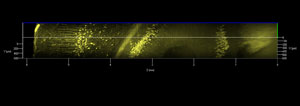 This enables 3-D visualization of the branches of individual nerve cells and imaging of their connections. In untreated tissue, penetration depth is now five to 10 times more than that achievable with a multiphoton microscope featuring traditional optics.
Using the microscope to visualize and track nerve cells deep into the brain is an important step in decoding brain circuitry. The new objective lens brings this goal closer to realization. The lens is designed for the clearing method known as “Scale,” developed by Dr. Atsushi Miyawaki at the Riken Brain Science Institute in Japan. A special water-based reagent solution transforms the sheath substance of the nerve cells into a transparent matrix without impairing the signals from fluorescent marker and tracer substances.
http://www.zeiss.com/microscopy
/Buyers_Guide/Carl_Zeiss_Microscopy_LLC/c16324
Published: February 2012
REQUEST INFO ABOUT THIS PRODUCT
* First Name:
* Last Name:
* Email Address:
* Company:
* Country:
Message:
When you click "Send Request", we will record and send your personal contact information to Carl Zeiss Microscopy LLC by email so they may respond directly. You also agree that Photonics Media may contact you with information related to this inquiry, and that you have read and accept our
Privacy Policy and
Terms and Conditions of Use.
Register or login to auto-populate this form:
Login
Register
* Required