
Three-Chip Color Camera
CANON MEDICAL COMPONENTS U.S.A., Video Sensing DevicesRequest Info
Toshiba Imaging Systems Div.’s IK-TF7 three-CCD progressive-scan color camera has been integrated into the Micron III retinal imaging microscope developed by Phoenix Research Labs.
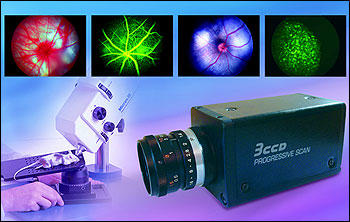
The system enables new modalities in high-resolution imaging for in vivo eye research, including white-light imaging of mice and rats, fluorescein angiography, diabetic retinopathy, retinoblastoma, choroidal neovascularization, retinitis pigmentosa and anterior segment slit-lamp. Live-animal fluorescent studies such as green and yellow fluorescent protein are also possible.
Camera features include 1024 × 768-pixel resolution, 4.65 × 4.65-µm pixel size and color-shading. Proprietary prism block color technology captures fast-moving items under test. The compact camera operates at up to 90 fps and eliminates image jitter via the ?-in. CCDs, whose co-site sampling arrangement eliminates RGB shift.
Other features include partial scanning, a field removable/replaceable infrared filter, on-screen and RS-232C setup, asynchronous reset, long-term integration and shutter speeds from 1/100 to 1/100,000 s.
https://mcu.canon/vsd
/Buyers_Guide/CANON_MEDICAL_COMPONENTS_USA_Video_Sensing/c15120
Published: March 2010
REQUEST INFO ABOUT THIS PRODUCT
* First Name:
* Last Name:
* Email Address:
* Company:
* Country:
Message:
When you click "Send Request", we will record and send your personal contact information to CANON MEDICAL COMPONENTS U.S.A., Video Sensing Devices by email so they may respond directly. You also agree that Photonics Media may contact you with information related to this inquiry, and that you have read and accept our
Privacy Policy and
Terms and Conditions of Use.
Register or login to auto-populate this form:
Login
Register
* Required