About This Webinar
Cellular metabolism is the process by which cells generate energy. The process can be dysregulated in many diseases and pathologies, including cancer, neurodegeneration, and diabetes. Current biochemical assays for metabolism are limited to either cell-destructive protocols, such as mRNA analysis, or they measure readouts from collective cell populations, such as oxygen consumption assays. Nondestructive measurements of metabolism with single-cell resolution are needed to study the contributions of metabolic heterogeneity to disease progression, cancer metastasis, and resistance to therapies.
Fluorescence lifetime imaging of the metabolic coenzymes, reduced nicotinamide adenine (phosphate) dinucleotide (NAD(P)H), and oxidized flavin adenine dinucleotide (FAD) provides a label-free method to interrogate cellular metabolism. Both coenzymes, NAD(P)H and FAD, exist in either a free or protein-bound configuration with a distinct fluorescence lifetime. Single-cell segmentation and analysis of fluorescence lifetime images allow metabolic measurements at the cellular level. To facilitate cell-level analysis of fluorescence images, Alex Walsh of Texas A&M University and her colleagues are developing automated segmentation algorithms. Additionally, they are creating and testing models for predicting cell phenotypes from fluorescence lifetime metrics.
Who should attend:
Researchers, clinicians, lab managers, engineers, and those who use fluorescence microscopy in their work. Those who are interested in or who work in imaging cells. Anyone who uses chemical microscopy, spectroscopy, photolysis, imaging, and molecular research in industries such as medicine, biomedicine, pharmaceuticals, test and measurement, and cancer research.
About the presenter:
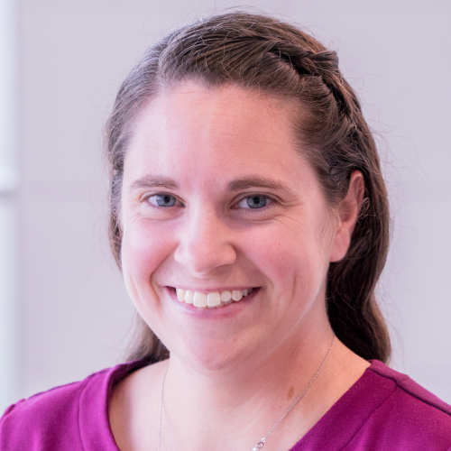 Alex Walsh (she/her) is an assistant professor in the Department of Biomedical Engineering at Texas A&M University. She leads the Quantitative Optical Imaging Lab, which seeks to develop and optimize optical imaging technologies for studying dynamic biological processes and identifying prognostic biomarkers of disease, progression, and response to treatment. Walsh is a founding member of her department’s Respect, Equity, Diversity and Inclusion Committee and works to increase diversity and inclusion in science and engineering.