About This Webinar

The microstructure and cellular organization of tissues have been imaged to differentiate between tissue types in their normal and diseased states since the 19th century. However, despite major medical advancements, histological examination of tissues still requires the tissues to be excised, processed, sliced, stained, and then imaged. Biopsy sampling removes valuable tissue, it may miss abnormalities, and it takes from 20 minutes to days to process — prolonging procedures that are driven by these biopsy results, driving up costs, and preventing closed-loop decision-making. MediSCAPE (swept confocally aligned planar excitation) is a single-objective light-sheet microscope that permits in situ, real-time three-dimensional histological imaging of living tissues without the need for excision. MediSCAPE’s ultrafast volumetric imaging speeds account for in vivo motion, and permit “roving” acquisition, which — when combined with 3D stitching — enables contiguous analysis of large areas or disease boundaries. MediSCAPE’s high sensitivity, even for weak intrinsic fluorescence, permits label-free, multispectral in situ imaging of clinically relevant tissue architecture without the need for exogenous staining, which could greatly facilitate clinical adoption.
***This presentation premiered during the 2022
BioPhotonics Conference. For more information on Photonics Media conferences, visit
events.photonics.com.
About the presenter
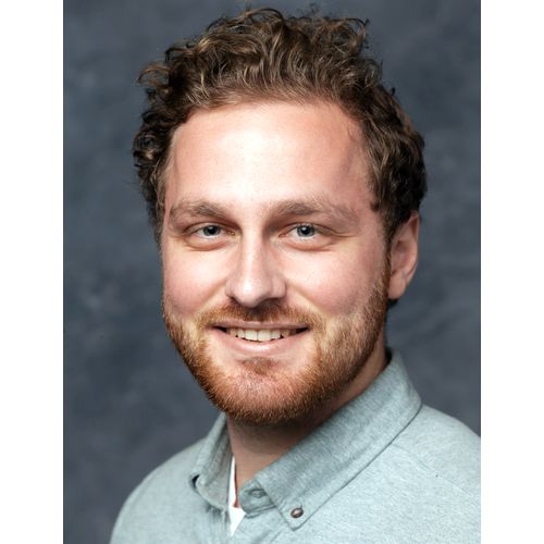
Malte Johannes Casper is a doctoral candidate at Elizabeth Hillman’s lab at Columbia University. He received a bachelor’s degree in medical computer science from the University of Lübeck in 2014 and a master’s degree in medical engineering science in 2017. During his master’s studies and beyond, he visited Massachusetts General Hospital in Boston, where he worked on the dermatological application of optical coherence tomography (OCT). He began his doctoral studies in 2018 at Columbia University, where he has been working on SCAPE and its application to large-tissue imaging in the clinical setting, as well as for neuroanatomical imaging.