HAYWARD, Calif., May 30 -- Researchers have demonstrated a new approach to using quantum dots for biological studies of living animals.
A new application of multiphoton microscopy makes tiny blood vessels beneath a mouse's skin appear so bright and vivid in high-resolution images that observers can see the vessel walls ripple with each heartbeat, 640 times a minute. The capillaries are illuminated in unprecedented detail using fluorescence imaging labels -- molecule-size nanocrystals called quantum dots -- circulating through the bloodstream.
Cornell University researchers, in collaboration with Hayward, Calif.-based Quantum Dot Corp., published their findings in the May 30 issue of the journal Science.
Watt W. Webb, a professor of engineering and applied physics at Cornell, is a co-inventor of multiphoton microscopy with Winfried Denk, and leader of the experimental imaging team at Cornell.
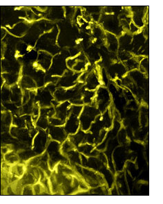
3-D VEINS: The branched capillary structure, feeding adipose tissue in a living mouse, is revealed with multiphoton fluorescence microscopy as nanocrystal quantum dots circulate through the bloodstream. Photo: Bioimaging Resource/Cornell University Copyright Cornell University
"Of course there are easier ways to take a mouse's pulse," said Webb's Cornell collaborator, senior research associate Warren R. Zipfel, "but this kind of resolution and high signal-to-noise illustrates how useful multiphoton microscopy with quantum dots can become, in a biological research context, for tracking cells and visualizing tissue structures deep inside living animals."
Zipfel cited the study of vascular changes in cancer tumors as one possible application. However, the Cornell researchers are not ready to recommend human-medicine clinical applications for quantum dot imaging, in part because some of the best fluorescing nanocrystals have unknown toxicity. Mice used in the Cornell study are still alive and apparently healthy, months later, and are being monitored for long-term effects of their treatments.
"We continue to be surprised at the broad array of biological applications enabled by our Qdot nanocrystals," said Marcel Bruchez, principal scientist at QDC and a coauthor of the Science article. This research will pave the way for many new noninvasive in-vivo imaging methods using quantum dots."
Quantum dots are nanometer-size semiconductor crystals that brightly light up specific biological events. These nanoscale molecular light-emitting diodes allow rapid, parallel, and ultrasensitive detection of biomolecular interactions.
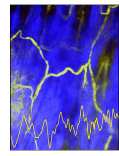
QUANTUM VIEW: Multiphoton fluorescence microscopy with quantum dots illuminates a capillary beneath the skin of a living mouse. In this image, collagen is imaged in blue by second harmonic generation; quantum dots inside the capillary are imaged in yellow by two-photon fluorescence excitation. Because red blood cells exlude the quantum dots, they appear as shadows within the capillaries, which can be monitored over time (yellow trace at bottom of image). Photo: Bioimaging Resource/Cornell University. Copyright Cornell University
Because of the special properties of the nanoparticles, multiphoton microscopy with quantum dot imaging can be a thousand times brighter in tissue than with conventional organic fluorophores (the chemical labels that are temporarily added to samples), said Webb. "We looked to quantum dots for even brighter images at better resolution, and that's what we found."
The results presented in the Science article show highly detailed images of capillaries beneath the skin of a living mouse after quantum dots were injected through a vein in its tail, as well as capillaries through the adipose (fat) layer around the mouse's ovaries. The researchers were particularly surprised at the saw-toothed ripples in the walls of one capillary image, until they made a calculation. Noting the time it took to scan that part of the tiny blood vessel and the animal's heart rate during the experiment, they determined that each ripple represented the undulation of the capillary wall from one heartbeat.
Besides demonstrating the feasibility of microscopic angiography with quantum-dot labeling through skin and adipose tissue -- two of the most challenging tissue types -- the researchers said they had resolved several fundamental questions, including the fact that sometimes as many as half the dots in a preparation are not fluorescent.
Other authors of the Science article are Marcel P. Bruchez, principal scientist at Quantum Dots; Rebecca M. Williams, a research associate with the National Institutes of Health (NIH)-funded Bioimaging Resource at Cornell; Frank Wise, professor of applied and engineering physics; and Stephen W. Clark, a graduate student in Wise's laboratory. Funding came from NIH, the Defense Advanced Research Projects Agency and the National Science Foundation.
For more information, visit: www.cornell.edu