Hank Hogan, Contributing Editor
Researchers have found dynamic force microscopy, or atomic force microscopy (AFM), useful for tracing surfaces on an atomic scale. The technique even may be capable of following biological samples in real time as they twist and turn. The difficulty has been in developing dynamic force microscopes with the right capabilities.
One problem has been building the devices. Another has been interpreting the results. Advances in fabrication techniques for micro-elecromechanical systems (MEMS) promise to solve the first. Now researchers at Kyoto University in Japan have come up with a solution to the second that could extend the application of dynamic force microscopy into new areas.
“It allows us to measure ultrahigh-speed phenomena with nanometer-scale resolution by force interaction, which can be microscopic biochemical mechanisms, molecular relaxation processes, device switching, magnetic spin processes and so on,” said Hirofumi Yamada, an associate professor in the university’s department of electronic science and engineering. He is a co-author of a paper outlining the approach that appeared in the Dec. 20 issue of Applied Physics Letters.
In dynamic force microscopy, investigators drag a miniature stylus across a sample. The stylus consists of a microscopic point attached to a cantilever a few tens of microns wide, and it deflects up and down as it travels across the sample. This response is measured by bouncing a laser beam at an angle off the cantilever. The beam acts as the arm of an optical lever, magnifying the minute motions of the cantilever into something more easily quantified.
The challenge is to follow those movements at high speed. Many commercial detectors used in AFM top out at approximately 1 MHz. The university researchers have beaten that limit using a heterodyne optical beam deflection method.
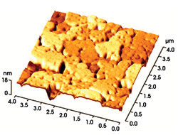
Using a heterodyne optical beam deflection method, researchers have investigated various surfaces, including Au(111), with high-speed dynamic force microscopy. Courtesy of Kyoto University.
The cantilevers vibrate like a diving board at a natural resonant frequency. In making a measurement with heterodyne optical beam deflection, investigators change the power of the probing laser beam at a rate close to this frequency. As a result, a beat forms, whose frequency is the difference between the pulsing of the laser beam and the vibration of the cantilever. Because it is at a lower frequency, the beat is more easily measured.
The scientists constructed 35-μm-long cantilevers with resonant frequencies of 5 to 8 MHz using a focused ion beam tool to sculpt the cantilevers and to deposit carbon tips on them. A Hitachi 5-mW, 635-nm laser diode, power modulated at a rate within a few tens of kilohertz of a given cantilever’s resonant frequency, probed the components. In doing so, they were able to measure various surfaces, including Au(111).
The researchers are trying to use the method to perform atomic- and molecular-scale imaging. They also would like to realize ultrahigh-speed measurements in the range of 100 to 1000 MHz. Achieving this will depend on developing cantilevers with the right characteristics.