WASHINGTON, Aug. 29 -- Researchers at Stanford University have demonstrated an optical technique that can capture micron-scale images from deep in the brains of live subjects. The method, called two-photon microendoscopy, combines a pair of powerful optical and mechanical techniques into a portable device based on advances in micromotors, lensing and fiber optics (fiber optic fluorescence imaging). The results will appear in the Sept. 1 issue of Optics Letters, a journal published by the Optical Society of America. The research team is led by Mark Schnitzer, an assistant professor with a joint appointment in Stanford's biological sciences and applied physics departments and a member of the faculty of the Stanford Brain Research Institute. Schnitzer has longstanding interests in neural circuit dynamics and optical imaging.
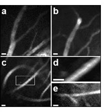
Images of cerebral blood vessels in the brains of living mice. The images show blood vessels near the surface of the brain, in the neocortex (panels a and b), and in the hippocampus (panels c-e) -- a brain area that is about 1 mm below the surface of the mouse brain. Images were captured 20 µms below the hippocampal surface (c and d) and 80 µms below the hippocampal surface (e). The scale (denoted by the white bars in each panel) is 10 µms, comparable to the size of individual nerve cells in the brain.
Obtaining images of living cells below the surface has been difficult with conventional techniques. Electron microscopy can't be used on living tissue, and optical (light) microscopy can't penetrate very deeply into tissue because light scatters as it travels through tissue near the surface. With one-photon fluorescence, the deep tissue causes photons to ricochet, or scatter, as they return to the detector, which creates a background haze in the images. With two-photon fluorescence imaging, instead of one higher-energy photon, researchers bombard a molecule with two photons of lower energy. Their combined energies total the energy required to excite the fluorescent-dye molecules used to mark the tissue. The technique gets rid of the background haze and reduces scattering, because molecules outside the area of interest are much less likely to absorb a pair of photons simultaneously and fluoresce in response.
Two-photon microscopy only penetrates brain tissue to about 500-600 microns -- barely scratching the surface. To get at the deep structures, the Stanford researchers turned to microendoscopy, using tiny, minimally invasive optical probes inserted deep into living brain tissue. To make one group of images (figures 1c-1e), the researchers inserted the microendoscope into the hippocampus, about a millimeter below the mouse brain surface. The two-photon imaging provided an additional 80 microns of depth, below the hippocampal surface.
The team used two-photon microendoscopy technique to capture detailed images of the blood vessels in the hippocampus sections of the brains of live mice. The mice were injected with a fluorescein dye, an FDA-approved contrast agent that is most commonly used for retinal exams in humans. The fluorescein labeled the blood plasma so the vessels in the brain could be clearly seen.
The Stanford researchers said the ability to take images of individual cells inside living subjects will provide insight into cellular behavior and how it affects organisms as a whole. For instance, the nerve cells of the hippocampus region contribute to important mental processes such as learning and memory, and certain deep-tissue areas of the brain are critical to understanding Alzheimer's and Parkinson's disease, for example.
For more information, visit: www.stanford.edu/