
Laser Provides New Insight into Human Brain
DURHAM, N.C., Sept. 26, 2006 -- In a finding that may offer clues about Parkinson's disease, a team of researchers used a sophisticated laser system and a photoelectron emission microscope to gain evidence that a dark brown pigment that accumulates in people's brains consists of layers of two other pigments commonly found in hair.
Other scientists previously had determined via chemical analysis that the dark pigment, called neuromelanin, is composed of the two pigments: eumelanin, found in black-haired people, and pheomelanin, found in redheads. But how those pigments are arranged structurally remained unknown -- and this structuring may prove to be of critical importance, according to the researchers.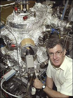
Duke University chemist John Simon with a photoelectron emission microscope at the laser lab. Simon and a team of researchers used the microscope and a sophisticated laser system to gain insights into the structure of pigments in the brain, a finding that could offer clues about Parkinson's disease. (Photo: Jim Wallace)
In addition, in 2005 a Duke University team that included some of the same scientists involved in the current study reported using the laser system to establish that pheomelanin is chemically disposed to activate oxygen while eumelanin is not. Oxygen activation is suspected to play a role in the neurogenic cascade of events behind Parkinson's disease.
In the new report, investigators from Duke, North Carolina State University and the Institute of Biomedical Technologies in Segrate, Italy, outlined evidence that neuromelanins isolated from human brains have cores of oxygen-activating pheomelanin covered by a protective surface of eumelanin.
"This is the first piece of morphological data about how these pigments are constructed," said study leader John Simon, the George B. Geller Professor of chemistry at Duke.
The findings "should stimulate renewed interest in the roles of neuromelanin in the pathogenesis of Parkinson's disease, the second most prevalent neurodegenerative disorder," Shosuke Ito, a chemist at Japan 's Fujita Health University School of Health Sciences, wrote in a companion commentary published with the study.
According to the team's report, neuromelanin granules begin appearing in human brains between ages 3 and 5, and their concentrations increase steadily thereafter. However, neuromelanin levels drop precipitously in the brains of Parkinson's patients, who also experience a death of brain cells that are darkly pigmented and an increase in brain tissue concentrations of the metal iron. Brain cells that produce dopamine, a key neurotransmitter disrupted in Parkinson's disease, experience high levels of oxidation as that dopamine is made, the researchers noted.
Scientists have hypothesized that brain cells synthesize neuromelanin to serve as a defense mechanism against high oxidation stress, the team's report said. Neuromelanin's layered granular structure could help protect brain cells from damage in several ways, Ito wrote in his commentary.
Having eumelanin at their surfaces would protect the granules with a pigment known to efficiently bind iron and other molecules that could otherwise play a role in oxidative damage. If the underlying core of pheomelanin were instead positioned at the surface, "the neuro-protective role of neuromelanin would not be expected," Ito added.
However, eumelanin is limited in how much iron it can take up, and other scientists have proposed that iron over-saturation at the granules' surfaces could contribute to the high levels of the metal in the brains of Parkinson's victims.
"Increased oxidative stress under such conditions could result in degradation of the eumelanic surface of neuromelanin," Ito wrote. That could expose a pheomelanin core "that is not only ineffective in iron-binding, but also behaves as a pro-oxidant itself," he added.
"Once these neuromelanin granules start getting chewed into, an environment is created that is much more pro-oxidation," Simon said. "As pigment starts to get eroded, you can imagine how oxidative stress could be increased in multiple ways."
In the study, the researchers used a special laser device that makes light with electrons that have been freed from their usual bondage to atoms. Housed in a large bay in the Duke University Free Electron Laser Laboratory, the device can be "tuned" step-by-step to produce light at a variety of different wavelengths, with each wavelength probing different energy regions in target molecules. The team also used a photoelectron emission microscope to resolve individual neuromelanin granules and distinguish between the two pigment types.
Using these devices in combination, the researchers could pinpoint the "oxidation potentials" of molecules coating the surfaces of neuromelanin granules. Oxidation potentials measure how likely given chemicals are to activate oxygen by giving up electrons. Activated oxygen can produce compounds called radicals that can stress cells.
The team found that oxidation potentials of molecules at the surfaces approximated those found in black hair pigments in the 2005 study. "That meant it was eumelanin, which is pretty antioxidant," Simon said. The laser beams could not penetrate beneath the granules' surfaces to record oxygen potentials nearer their cores. But previous chemical analyses by other researchers had established that neuromelanin is a mixture of both red and black hair pigments. So, the new finding suggests "a structural motif, with pheomelanin at the core and eumelanin at the surface," the team reported.
"Something special is happening, where the red pigment is getting encased in the black," Simon said. "So the red, being fairly pro-oxidant, is being encased in this antioxidant pigment."
Simon's group could only deduce the probable structure of neuromelanin, rather than measure it directly, because scientists have so far been unable to synthesize the pigment from chemical building blocks in a form that duplicates the natural version, he said.
"No one knew how to test or probe these things," Simon said. "And I can't overestimate how difficult it was to get materials to test." His group worked with small amounts of autopsied brain tissues provided by a research group led by Luigi Zecca at the Italian Institute of Biomedical Technologies.
First author of the study is Simon's graduate student, William Bush. Other researchers working on the study were Glenn Edwards, director of the Duke University Free Electron Laser Laboratory; Robert Nemanich and Jacob Garguilo of NC State and Fabio Zucca and Alberto Albertini of the Italian Institute of Biomedical Technologies.
The team published the findings online this week in the journal Proceedings of the National Academy of Sciences. The research was funded by the US Air Force Office of Scientific Research through grants to the Duke University Free Electron Laser Laboratory, and by the Italian Fund for Basic Science. For more information, visit: http://dukenews.duke.edu
Published: September 2006