A new imaging method has emerged that allows scientists to see cell molecules more clearly.
Niels de Jonge and colleagues at Vanderbilt University and Oak Ridge National Laboratory (ORNL) have discovered a technique for imaging whole cells in liquid with a scanning transmission electron microscope.
 “Electron microscopy is the most important tool for imaging objects at the nano-scale – the size of molecules and objects in cells,” said de Jonge, an assistant professor of molecular biology and biophysics at Vanderbilt and a staff scientist at ORNL. But electron microscopy requires a high vacuum, which has prevented imaging of samples in liquid, such as biological cells.
The technique, dubbed Liquid Stem, uses a microfluidic device with electron transparent windows to enable the imaging of cells in liquid. The team has demonstrated the imaging of individual molecules in a cell, with significantly improved resolution (the fineness of detail in the image) and speed compared with existing imaging methods.
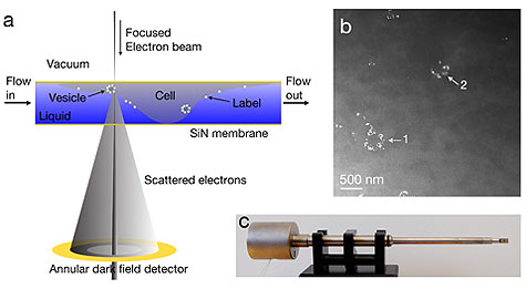
(a) The principle of liquid scanning transmission electron microscopy (Liquid Stem) is shown. A whole cell in liquid is enclosed between two electron-transparent windows of silicon nitride. Images are obtained by scanning a focused electron beam over the sample and detecting the elastically scattered electrons with an annular dark field detector. Labels made of a high atomic number material can be distinguished. (b) Liquid Stem images of COS7 fibroblast cells incubated with 10-nm gold nanoparticles conjugated with epidermal growth factor (EGF-Au) are shown. The labels are visible as bright spots and the cellular material is shown as light-gray matter. Sharp-edged gold labels are visible in the cluster at arrow #1, while the labels in the cluster at arrow #2 appear blurred and cannot be distinguished as individual labels, indicating that cluster #2 is out of the vertical plane of focus.The observation of circular clusters of labels at different vertical positions in the cell indicates that the labels were internalized. The clustering of the EGF receptors and internalization of the labels is consistent with the known behavior of EGF-activation of EGF receptors, which cluster as internalized endosomes upon receptor activation. (c) The flow cell is placed in the vacuum of the microscope using the fluid specimen holder shown here. Image courtesy of PNAS.

The technique also could become a resource for energy science, as researchers use it to visualize processes that occur at liquid; for example, solid interfaces in lithium ion batteries, fuel cells or catalytic reactions.
“Our key innovation with respect to other techniques for imaging in liquid is the combination of a large volume that will accommodate whole cells, a resolution of a few nanometers and fast imaging of a few seconds per image,” de Jonge said.
The research was supported by the Laboratory Directed Research and Development Program of ORNL, the SHaRE User Facility at ORNL, Vanderbilt University Medical Center and the National Institutes of Health.
For more information, visit: www.mc.vanderbilt.edu