In research hospitals and in the pharmaceutical industry, pathologists have been using special scanners to record digital images of tissue slides. Now these digital pathology scanners are expanding from research markets into what is projected to become a $2 billion to $4 billion industry.
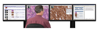
Aperio makes a picture archiving communications system, or PACS, which is essentially a command center that enables pathologists to review digital slides.
“There seems to be a lot more interest than there was two years ago,” said Anil Vasdev Parwani, MD, PhD, director of the division of pathology informatics at the University of Pittsburgh Medical Center, a leading research institution in the field of digital pathology.
Digital pathology systems can capture images of entire slides with resolution approaching that of a microscope. However, this was not the case until recently. Originally, digital pathology systems consisted essentially of a microscope and a digital camera, which is why digital pathology sometimes is called virtual microscopy.
With time, digital pathology systems broke away from the microscope and camera model and began incorporating nonstandard optics, illumination and sensors. Computers became faster, and computer memory increased. Today, some digital pathology systems are automated and even include a command center for the pathologist that often is referred to as a picture archiving communications system, or PACS.
Perhaps the only remaining technical limitation is that the images occupy gigabytes of computer memory. Besides taking up space, the large image files can slow down computer and network processes. “We have made a lot of strides in that area, but a lot of work needs to be done,” Parwani said.
Competition increases
The chief advantage of digital images over glass slides is that digital images can be accessed anywhere. “If you’re a pathologist in Cleveland and you want to consult a colleague in California, you can do it,” Parwani said. Pathologists also can share images with colleagues worldwide, not only for consultations but also for training purposes. And digital images can be read with software programs that help pathologists make more accurate diagnoses.
Before digital imaging, pathology labs had to ship glass slides, which can break or get lost during transit and which sometimes are not stored in a convenient place. For example, a pathologist on the fifth floor of a hospital may want access to slides that are in the basement. With digital pathology, the doctor can download images of the slides right there.
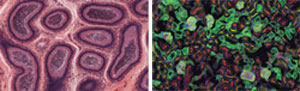
But is it art? This fluorescence image was taken with an Aperio system.
However, not all pathologists have a positive attitude toward the technology. “The initial reaction is one of fear; they think it’s going to replace them,” said Dirk Soenksen, founder and CEO of the market-leading digital pathology company Aperio Technologies Inc. of Vista, Calif.
Although the technology could enable outsourcing to underpaid medical professionals in developing countries such as India and China, both Parwani and Soenksen believe that the technology instead will drive cases to the US, where there is a greater concentration of expertise.
Despite some misgivings about the technology, its potential has generated considerable excitement – which is why General Electric Co. has entered the fray. The corporate giant’s health care business unit, GE Healthcare, recently joined forces with the University of Pittsburgh Medical Center to form the digital pathology venture Omnyx LLC, with offices in Pittsburgh and Piscataway, N.J.
From the sound of its chief, Aperio isn’t running scared at the thought of GE entering the marketplace. “I think the question is: How does Omnyx plan to compete with us?” Soenksen said. He was quick to point out that Aperio has 500 systems in 44 countries and several FDA clearances, whereas Omnyx has yet to develop and sell a single product.
On the other hand, Soenksen, who once worked for GE himself, had praise for the people behind Omnyx, calling them “very smart people.” He said that the recent foray of GE into the digital pathology marketplace was actually a good thing because it has convinced financial firms of the value of digital pathology. Aperio already has raised $53 million in venture capital, including $20 million that it raised during the 2008 recession year.
Aperio isn’t the only player in the digital pathology market. Others include BioImagene Inc. of Cupertino, Calif., and the microscopy companies Carl Zeiss, Olympus, Nikon and Leica. However, Soenksen said that Aperio is the only one to offer every type of digital pathology product, including slide scanning and storage services for customers who would rather pay for those services than buy the equipment.
Software teaches doctors a thing or two
The life sciences sector of Definiens specializes in digital pathology software. According to Manfred Voglmaier, vice president of business development for that division, Nobel laureate Gerd Binnig founded the company upon the insistence of a journalist with the German newsmagazine “Stern” (in English: “star”).
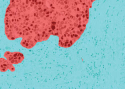
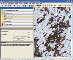
Aperio’s software can differentiate tissue types by recognizing patterns, as shown here.
From these beginnings, Definiens created TissueMap software, which identifies and relates objects on pathology slides in a way that is designed to be intuitive. The software is marketed at pharmaceutical and biotechnology firms that analyze tissue slides during the drug development process.
Although several other companies make similar products, Voglmaier contends that TissueMap is unique because it uses context-based rule sets to automatically identify and quantify various cell types and subcellular structures. Besides the standard rule sets, customers can use those they have made themselves, and the company can make rule sets upon request. The company holds more than 30 patents on the software.
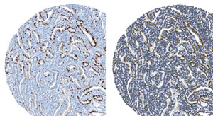
Definiens TissueMap software classified positive (yellow) and negative (blue) nuclei in this tissue microarray core by means of Ki67 staining. Courtesy of sanofi-aventis.
In a proof-of-concept study of the software for Big Pharma, pathologists reviewed 1800 slides without the software. Concurrently, the software reviewed the same slides without any input from the pathologists. Surprisingly, the TissueMap software correctly labeled 30 percent more of the slides than did the pathologists.
Shown is a TissueMap software analysis of antibodies and markers that co-stain the cell body.
Currently, the software is for research only. In several months, the company plans to sponsor clinical trials to obtain FDA clearances. The latest version of the software, 3.0, has enhancements that include support for tissue microarrays.
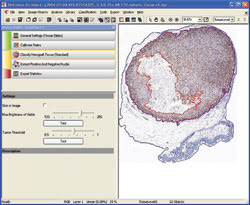
TissueMap software offers fully automated detection of a tumor graft and separation of living and dead regions of tissue.