
Donors and Acceptors: New Developments in FRET
Förster resonance energy transfer, or FRET, has contributed to advances in a host of applications. Measuring the energy transfer between donor and acceptor dyes attached to two ends of a molecule, or to two different molecules brought close together, enables researchers to probe a range of biological processes, including protein-protein and protein-DNA interactions, ligand-receptor binding, and protein conformational changes in response to a biological stimulus. It also allows them to explore spatial relationships in biological structures and macromolecules by monitoring the distance between two fluorophore-labeled sites.
FRET microscopy has proved a popular means of characterizing molecular interactions in living organisms. Also, this intermolecular sensing capacity facilitates a variety of bioanalytical and biosensing applications.
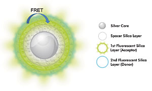
Researchers have reported a FRET technique that uses multilayer core-shell nanoparticles to exploit the phenomenon known as metal-enhanced fluorescence. Shown here is a schematic representation of metal-enhanced FRET in the nanoparticles. The silver core is surrounded by concentric layers of fluorophore molecules, and the distance between them is precisely adjusted by adding a silica spacer shell with controlled thickness. Coupling of the core with the fluorophore molecules increases the distance over which FRET occurs, and fluorescence intensity is significantly enhanced.
Development of the technique is ongoing. In recent years, FRET has benefited from a variety of new components and methodologies. Improvements have been made possible, said Sapna K. Deo, assistant professor of bioanalytical chemistry at Indiana University-Purdue University Indianapolis, by the availability of inexpensive powerful lasers and detectors such as avalanche photodiodes and photon multiplier tubes, by the evolution of fluorescence microscopy, and by the development of better filters, better software for data analysis and better cameras.
Use of novel donors and acceptors has added further edge to its development, she said. Conventional fluorophores used with FRET - including organic dyes, fluorescent proteins, chemiluminescent substrates and fluorescent polymers - often are inherently limited. Their narrow excitation windows and broad emissions can result in spectral overlap between the donor and acceptor, for example, which can cause headaches, especially when monitoring interactions between two or more donor-acceptor pairs with multiplexed FRET. Other concerns are limited photostability and the possibility of photobleaching and photodegradation. Researchers have explored several alternatives to sidestep these issues. Recent years have seen the development of metal nanoparticles and bioluminescence for FRET enhancement. Each of these offers a unique set of advantages with respect to conventional fluorophores and thus could advance the technique for a wide array of applications.
Metal-enhanced fluorescence
In Québec City, researchers have achieved FRET enhancement using multilayer core-shell nanoparticles to take advantage of a phenomenon known as metal-enhanced fluorescence. Placing donor-acceptor pairs around or near metal nanoparticles results in coupling between the local electric field from the excitation source, the electrons in the metal and the electric dipole induced in the dye molecules, increasing the strength of the donor-acceptor interactions and thus improving FRET efficiency.
Denis Boudreau, professor of analytical chemistry and a member of the Centre for Optics, Photonics and Lasers at Laval University in Québec City, understood that the efficiency of this approach is strongly dependent on the separation between metal and fluorophore and, therefore, that investigators could maximize it by gaining precise control over this distance. To this end, in a Nano Letters paper published online on July 15, he and others in his lab - doctoral student Mathieu Lessard-Viger, aided by graduate students Maxime Rioux and Luc Rainville - reported a study in which they fabricated core-shell nanoparticles and chemically grafted fluorophore molecules within concentric silica shells so they could adjust at will the distance between fluorophore and metal to optimize FRET enhancement.
These novel luminescent nanoparticles provide two important advantages, Boudreau said. First, they yield significant increases in fluorescence intensity. The researchers reported a fifteenfold increase with the particles used in the Nano Letters study, and they anticipate even higher increases with thinner spacer shells in their design. More importantly, use of the nanoparticles resulted in significantly enhanced FRET.
Boudreau and colleagues calculated an energy transfer efficiency between donor and acceptor of 56 percent, as opposed to an efficiency of only 12 percent for nanoparticles in which the metal core was etched away. Furthermore, they noted that the Förster distance - the range at which the energy transfer efficiency is 50 percent - was 30 percent higher than that published for a fluorescein-eosin system.
This increase in Förster distance recommends the nanoparticles for a variety of imaging microscopy and biosensing applications, Boudreau explained; for example, by conjugating them to biomolecules and using them as longer-range donor-acceptor pairs.
The researchers continue to develop the nanoparticles with an eye toward these and other applications, including ultrasensitive DNA detection. They also plan to adapt them to multiplexed signaling by varying their dyes and relative concentrations and by tuning their emission signatures.
Bioluminescence resonance energy transfer
Bioluminescence resonance energy transfer, or BRET, represents another important advance in the field. The technique, which uses a bioluminescent donor and a fluorescent acceptor, overcomes many of the obstacles associated with FRET-based methods. First, because it does not require an external source for excitation, as do the latter methods, it is less expensive and offers improved efficacy for in vivo applications. Also, it does not have the same issues as FRET with photobleaching and simultaneous excitation of donor and acceptor fluorophores and, as a result, offers significantly reduced background noise. Finally, it is more compatible with photoresponsive tissues than are FRET-based methods.
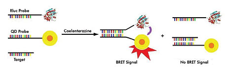
Scientists have described a bioluminescence resonance energy transfer technique in which Renilla luciferase is used as a bioluminescent donor and quantum dots as acceptors. The system described employs two probes: one conjugated to Renilla luciferase and complementary to the target sequence (Rluc probe in this figure), the other conjugated to a quantum dot and homologous to the target sequence (target probe). When the target nucleic acid is not present, the two probes hybridize, bringing the luciferase and quantum dot nearer to one another and thus producing a BRET signal.
Deo's lab focuses on development of BRET-based methods for quantitating microRNAs. MicroRNAs play an important role in gene regulation, she said, and thus have attracted attention both clinically as disease biomarkers and in fundamental biomedical investigations. Researchers in the lab have developed a BRET-based technique that employs Renilla luciferase as a bioluminescent donor and quantum dots as acceptors, and they have described its use to detect DNA. The BRET system reported uses two linear oligonucleotide probes. The first, conjugated to Renilla luciferase, is complementary to the target sequence. The other, conjugated to a quantum dot, is homologous to the target sequence. The two probes hybridize when the target nucleic acid is not present, bringing the luciferase and quantum dot closer together and producing a BRET signal.
When the target nucleic acid is present, however, it competes with the luciferase probe for hybridization of the quantum dot probe. This results in a decrease in the BRET signal and an increase in the luciferase emission, enabling researchers to measure the nucleic acid.
To quantitate the DNA, the researchers plotted a calibration curve of BRET ratio vs. target concentration. They calculated the BRET ratio as bioluminescence intensity at 485 nm divided by bioluminescence intensity at 705 nm. Based on the resulting ratio, they quantitatively determined the amount of DNA target from the equation of the calibration curve.
The researchers continue to develop the technique and are working toward multiplexed detection of target nucleic acids in the solution phase. This can be accomplished using multiple spectrally distinct quantum dots. Because of their overlapping absorbance spectra, multiple quantum dots can be excited simultaneously with a single source, such as Renilla luciferase.
"Therefore," Deo said, "utilizing Renilla luciferase as BRET donors and multiple quantum dots as BRET acceptors, multiplex detection can be achieved. The DNA sequences of the oligonucleotide probes need only be altered to specifically hybridize with their respective targets."
Also see: More Web Exclusives
Published: September 2009