
Making Medical Devices and, Hopefully, Money
To apply an old saying to the commercialization of research, there’s many a slip before a product ships. Turning something developed in an academic setting into a viable medical device that can be mass-produced while hitting cost, reliability and performance targets is no easy task. In the end, turning out a successful product may depend as much upon the researcher as it does the research. A look at a couple of cases illustrates this point.
Miniature advance, massive payoff
At privately held startup EndoRobotics Inc. of New York, plans are under way to commercialize a medical device developed at, and licensed from, Columbia University, also in New York. Dubbed VisionTrackerOne, it is a fully insertable robotic imaging and surgical device for minimally invasive procedures.
The origins of the tool are important, said president and CEO James Wylie. “What you have is a leading surgeon’s viewpoint of what is needed to accelerate the conversion of open-cavity surgeries to minimally invasive surgeries.”
The Columbia research team included Dr. Dennis Fowler and Peter Allen. The former is a pioneering laparoscopic surgeon, while the latter is a computer science professor. With their backgrounds, they had firsthand experience with the shortfalls of current instruments, along with knowledge of what’s now technically possible.
Fowler, who is now chief medical officer at EndoRobotics, said traditional laparoscopic imaging involves inserting a glass rod into the patient, with the rod conducting light to an external camera. The rod is kept in place for the duration of the procedure. Consequently, it takes up room and prevents the opening from being used by anything else. Moreover, the imaging is two-dimensional, but the space being looked at occupies three dimensions.
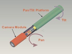
This device, which is in the process of being developed by EndoRobotics, is designed to be inserted into a patient, providing surgeons with the 3-D imaging needed for a better view during minimally invasive surgery. Courtesy of EndoRobotics.
A final problem is that navigating that space with the imager is counterintuitive. “To look to the right, you have to move your hand controlling the device to the left and vice versa,” Fowler said. “You have to move up to look down and vice versa.”
The new device, which is possible only because of advances in sensors and actuators, eliminates many of these problems. Consisting of a 110 × 10-mm fully insertable modular camera with an integrated lighting system, it can pan, tilt and zoom. Control is either by joystick or automatic surgical instrument tracking. It can have one or two sensors, with the latter configuration enabling stereoscopic imaging. The initial implementation, expected in 2011, will be 2-D, with a 3-D version deployed soon after.
Technological changes may alter some of these plans. For example, the initial device will be reusable, built to withstand repeated autoclave sterilization and the associated handling. However, advances in CMOS sensor quality and cost make a disposable device a possibility, which would eliminate the need for multiple sterilizations and potentially lead to significant cost reductions for hospitals and surgical centers.
As with many medical devices, regulatory approval is required. Wylie said that the device should be in the Class II category, with similar devices having preceded it and having already been approved. If that’s the case, it should move fairly easily through the approval process. Manufacturing the device in volume will be done by a third party. According to Wylie, the manufacturing partner will be selected after EndoRobotics completes its initial financing round.
As for the payoff, projections from the company are for revenues of more than $110 million four years after product launch. Those figures depend largely on an expansion of minimally invasive procedures currently performed by laparoscopy as well as on a certain percentage of open-cavity surgeries switching to the laparoscopic approach.
Getting a better view
A similar focus on solving an existing problem for physicians is behind a device being commercialized by Vital View, a startup in Misgav, Israel. Dr. Lior Yankelson, the company’s director and head of business development at the Misgav Venture Accelerator, said that the enabling technology involves microfiber optics. These have seen significant improvements over the past five years, particularly in commercially available products.
“The technology has not only matured but has come to a stage where it’s really advanced. The materials today are very robust and very durable, with relatively low cost,” Yankelson said.
That has made it possible to put the fibers in very small-diameter devices. The result has been that it’s now feasible to capture images in places where before it was not possible.
As for the problem being solved, that involves the delivery of an embryo, via catheter, to the womb during in vitro fertilization. To do this, physicians navigate the catheter through the patient’s cervical canal, a process that today depends upon feel because there’s no good way to see clearly in the liquid, secretion-rich environment.
Commercializing technology originally developed at Columbia to address this issue, Vital View hopes to have its first prototype for clinical use by the end of this year. Physicians will be able to see the catheter’s surroundings on a screen, making the embryo transfer process potentially both easier and more successful. Yankelson stressed that nothing about the procedure will change, except for the introduction of a visualization method.
He noted that millions of in vitro fertilization procedures are done worldwide every year, and Israel is one of the largest markets. The hope is that regulatory approval will be relatively quick since similar devices have already been approved and are on the market. If so, then significant amounts of data on the safety of those devices are potentially already available.
It also means that no clinical trial may be needed for FDA approval. In that case, Vital View would still conduct clinical trials, doing so to prove the efficacy of the device, Yankelson said.
An important point is that the original research and development of the device was done by Gary Nakhuda, a practicing reproductive specialist at Columbia. That means that the inventor knew what the problem was and how the solution addresses those issues.
That fits the template of the type of research sought by some investors, such as the Israeli business group to which Yankelson belongs. “We very much like products that are made by practicing physicians.”
Published: September 2009