Advances in technology are making digital microscopy more useful for education, research and clinical applications.
Palos Pathology Associates
Ltd. provides pathology services to a 300-bed community hospital in suburban
Chicago, and Dr. Stephen G. Ruby, president of the company, is thinking of expanding
the geographic area that it covers. In the past, that would have meant persuading
clinicians practicing at remote sites to make a trek to the hospital if they wanted
to review prepared slides with a pathologist. Now, the company has a system that
scans a physical slide and turns it into a virtual one that can be viewed over an
Internet connection.
A pathologist can scan a slide, note an area of
interest and e-mail a link to a clinician. “When they open it, it will open
to that exact spot that I’m showing them, but yet they will still be able
to then move around and look at the rest of the slide if they want,” Ruby
said.
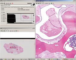
Virtual slide software converts pathology
slides into virtual images that can be viewed remotely and stored as digital data.
This screen shot of the output from a Nikon Coolscope VS was rendered by software
from Bacus Laboratories. On the right is the image of a pathology slide that would
be seen through a microscope, while on the bottom left is a macro view, showing
the location of that close-up. Above that are software controls. Courtesy of Palos
Pathology Associates.
What’s more, the two could look
at the same slide over the Internet while talking on the phone. The experience,
he said, would be similar to sitting at a two-headed microscope in the lab.
Palos is using a Nikon Coolscope VS,
which was acquired about a year and a half ago. Ruby would like it to scan faster,
but he’s happy with the system for the most part — especially the visual
quality.
The system combines Nikon’s microscope
optics and five-megapixel digital camera with software from Lombard, Ill.-based
Bacus Laboratories Inc. Ron Zibilich, clinical sales manager for Nikon Instruments
Inc. of Melville, N.Y., noted that the software helps keep the bandwidth needs of
the device low. The system moves about 70 kB of imaging data over the connection
at a time, which makes viewing of images much easier and much faster.
There are other virtual slide systems,
including the Bliss system from Bacus, the NanoZoomer Digital Pathology System from
Japan’s Hamamatsu Photonics KK, the Mirax Scan from Germany’s Carl
Zeiss MicroImaging GmbH, and .slide from Soft Imaging Net, also in Germany. Of these,
the last isn’t available in the US.
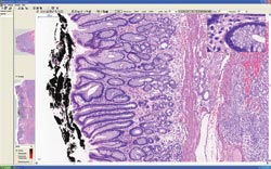
Here a slide of gut tissue
as displayed with Mirax Viewer virtual slide software. On the left of the screen
shot are two overview images of different magnification that show the position of
the high magnification image on the right.
These products all have objectives
and controlling computers, along with image storage systems. But they differ in
various ways. The NanoZoomer, for example, uses a 4096 x 64-pixel CCD sensor that’s
moved across the real slide to create the virtual one.
The Coolscope, Bliss, Mirax and .slide
systems, in contrast, employ more conventional cameras. All the systems stitch separate
images together to make a single virtual slide composed of up to billions of pixels.
Bacus sells the Coolscope, Bliss and
NanoZoomer in the US. Amanda J. Lowe, national sales manager for Bacus, said that
the three systems vary in cost and intended markets. The Coolscope costs about $25,000
and is a single-slide system intended for education and low volume settings. Virtual
microscopy systems are useful in educational settings because medical and other
students need to view slides but do not need to be trained in general microscopy.
So a virtual slide can be a good learning tool, especially because the slide
set won’t get lost or broken and won’t degrade over time. Students can
view these virtual slides wherever there’s an Internet connection with adequate
bandwidth.
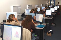
One of the uses for virtual slide systems is in teaching. Students
can all view the same slides, without the problems of variation, damage
or degradation that are found in physical slides. Students can also
access the slide images when outside of the classroom, including the
local coffeehouse — provided that they have an Internet connection with
enough bandwidth. Courtesy of Bacus Laboratories.
The Bliss system is more expensive
and faster, and it has more options for objectives and possible customization. Useful
in research applications, it saves images in a digital format without requiring
pictures to be taken manually or semiautomatically with a camera.
On the high end is the NanoZoomer, which tops out at well over $100,000. It has a 210-slide loader and can scan a slide at 20x magnification in about four minutes per slide at 20x and 2 1/2 times as long at 40x. The primary markets for this product are those that demand a high volume of slides for capturing such as pharmaceutical, clinical and research laboratories, Lowe said. In clinical settings, a virtual slide can be attached to
patient records so that the information doesn’t get lost and the images are
readily available for a consultation.
Gigabytes and megapixels
Although a onetime skeptic, Harvard Medical School
pathologist Wolfgang Klietmann is convinced that virtual slide and digital pathology
technology now comes with a quality and at a cost that make it useful. The instruments
deliver the resolution of digital photography and can be used over the Internet.
Improvements in bandwidth allow those images to be transported across the room
or around the world without a bothersome delay, he said.
He thinks this type of microscope will
gain use in many medical disciplines because it will help those working in pathology
interact with those in internal medicine, surgery, hematology, pediatrics and other
clinical areas. “Digital pathology will also improve medical care by making
more expertise available to remote places and decrease medical errors and lower
cost,” he predicted.
Klietmann is working with the start-up
company Corista LLC, which is developing a turnkey digital pathology solution. The
company is using a Zeiss system as a basis for its product, largely because of the
quality of its optics and electronics.
There is still a problem, though, with
file size. The data in an image depends upon the area of the slide and the magnification
of the scan. Systems that capture data in stacks that span the entire depth of a
specimen can add a third dimension.
Klietmann said that some of the files
can top 10 GB, making them difficult to manipulate and store. It also could cause
network congestion, particularly if several users are attempting to view virtual
slides at once. More bandwidth could overcome this problem, but it may be necessary
to employ lossless compression algorithms that reduce the size of image files without
compromising the data.
Improvements have made handling such
large files less of a problem, said Norbert Schuster, product marketing manager
for imaging systems at Carl Zeiss MicroImaging Inc., the US subsidiary in Thornwood,
N.Y. For example, improvements in storage media (in particular, DVDs), the advent
of bigger hard drives, falling server prices and development of affordable high-resolution
displays are all helping make virtual slides and digital pathology possible.
The Zeiss system, he explained, is
intended for high-volume applications where someone might load many slides, start
the system and run it overnight. It can hold 300 slides, taking 10 minutes to scan
one that measures 15 mm2 at 20x magnification. The resolution, determined by the
optics, is 0.23 μm per pixel.
Besides market forces, Schuster also
sees regulatory pressure playing a role in the adoption of virtual slide technology.
There’s an increasing emphasis on digital records, and regulators are pushing
hospitals and clinics to adopt such technology. As a result, he foresees significant
growth in sales of the virtual slides. “I think two to three times higher
growth rates over the next year or two as compared to the conventional microscope
market,” he said.
X-ray vision
For pathology, radiology’s transition from
film to digital might hold clues about a likely future as well as possible problems.
More than a decade ago, radiologists began the transition. Along the way, they have
hammered out standards and developed an infrastructure to handle such matters as
retrieving images on command and ensuring that images weren’t altered.
For example, digital radiology has
computer-aided diagnostics — software that helps pinpoint potential problem
areas. There’s a possibility that pathology packages could someday offer the
same capabilities, with enhancements such as automatic measurement and quantification
of features likely to be available much sooner.
Cameila D. Johns, a pathologist on
the faculty of the University of Tennessee Health Science Center in Memphis, is
interested in the telepathology aspects of these systems (see “Pathology at
a Distance”). She noted that the technology for virtual slides already exists
but that some of the business issues still have to be worked out.
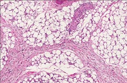
This virtual slide sample of human tissue was fixed in formalin and embedded in paraffin
before being cut on a microtome. It was then stained in a hematoxilin-eosin solution,
mounted on a glass slide, and finally covered with a thin glass coverslip. After
being prepared, its image was captured on a turnkey digital pathology system based
on a Zeiss microscope and a camera being developed by Corista LLC.
For example, who pays for a primary
virtual slide diagnosis? Although this isn’t being done, it is a potential
for certain types of tissue. The coding for insurance, reimbursement rates, procedures
and other business aspects are well established for actual glass slides and any
associate pathology. It isn’t clear whether looking at a digital slide can
be billed or reimbursed in exactly the same manner as is done when looking at a
glass slide. “That’s one of our hurdles now, to make this something
which would be recognized as equivalent to a slide diagnosis,” Johns said.
Beyond that, there’s the question
of FDA approval, which will be needed for certain applications in the US. At present,
none of the systems has the regulatory agency’s authorization for use in clinical
diagnostics. For now, virtual slide suppliers are concentrating on other markets
in the US, such as research and teaching applications, that don’t have these
regulatory requirements.
As for end users, they aren’t
using such systems for primary clinical diagnostics, either. Ruby of Palos Pathology,
for example, uses his setup to consult with physicians for diagnostic purposes,
likening this to an adjunct to a telephone conversation.
Gaining the FDA’s OK will require
validation studies comparing the technology with the old standard, and such studies
will have to be done for each type of system. Virtual-slide vendors haven’t
announced when — or even if — they’ll undertake these validation
studies. However, some of those who plan to market systems aimed at diagnostic settings
are working on validating the technology to the degree needed.
Although such business issues are being
resolved, potential virtual slide system customers are faced with some choices.
A number of commercial systems are available, all with different capabilities, price
points and intended targets. Choosing among them can be difficult.
Ruby emphasized that end users must
know what they want to do with a system before they buy it. That means an honest
and complete assessment of needs, and then due diligence when looking at all aspects
of a product to make sure that it fits present and future needs.“You can’t
just go in and buy something and say, ‘I’m going to work around what
it can do,’” he said.
Pathology at a Distance
The most important aspect of several available virtual slide systems is that they also offer
telepathology functions, according to Dr.?Cameila D. Johns of the University
of Tennessee in Memphis. She is director of the Path Connection, a nonprofit endeavor
by the university’s Health Science Center that specifically looks at difficult
cases in underserved populations. The group does this through real-time digital
imaging, with volunteer pathologists operating the instruments remotely so that
their expertise can be put to use at a distance.
Those distances can be impressive.
One of Path’s programs involves working with a hospital in Malaysia, and there
are plans to open another site in Cameroon. Because of Malaysia’s location
relative to Tennessee, the telepathology is done not only across space, but also
across time zones.
“Most of the cases that we are
looking at happen actually after working hours in the US. So it’s 8 o’clock
in the night when I’m actually looking at cases,” Johns said. She does
the initial slide screening and then consults with other pathologists.
Her group uses the Coolscope from Nikon,
and because the instrument sits at an Internet address, anyone with a browser can
navigate to it, provided that they satisfy any security requirements. She noted
that it typically needs only a 512-kb/s connection to make the instrument function
and that that relatively low requirement is important, given the areas it intends
to reach with telepathology.