Compiled by Photonics Spectra staff
A new nanoscopic imaging technique may lead to experimental methods for early detection and diagnosis of – and possible treatments for –pathological tissues that are precursors to multiple sclerosis (MS) and similar diseases.
Chemical engineers at the University of California have studied the myelin sheath, the membrane surrounding nerves that is compromised in patients with ms. Their findings appeared in the May 23, 2011, issue of Proceedings of the National Academy of Sciences (doi: 10.1073/pnas. 1106368108).
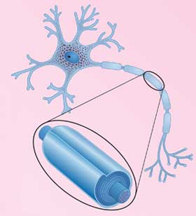
Shown is an illustration of a neuron, the basic functional unit of the nervous system. The enlarged detail indicates the myelin sheath, a multilamellar membrane surrounding the axon of neurons that is damaged in multiple sclerosis patients. Courtesy of Dottie McLaren.
Various parts of the central nervous system, including the brain, communicate with each other throughout the body via the transmission of electric impulses along the fibrous myelin sheaths, which act like electric cables or transmission lines. Defects in the molecular or structural organization of the myelin membranes lead to a reduction in transmission efficiency, resulting in sensory and motor disorders or disabilities – and neurological diseases, including multiple sclerosis.
MS is characterized by the appearance of lesions or vacuoles in the myelin at both the micro- and macroscopic levels, which results in the complete disintegration of the myelin sheath in a process called demyelination.
The paper describes fluorescence imaging and other measurements of domains, small heterogeneous clusters of lipid molecules – the main constituents of myelin membranes – that are likely to be responsible for the formation of lesions. The scientists conducted their research using model molecular layers in compositions that mimicked both healthy and diseased myelin membranes.

Fluorescence images of lipid domains in model myelin monolayers show coexistence of liquid-ordered (dark) and liquid-disordered (pseudocolored) phases. Depending upon the conditions, the lipid domains can exist in various shapes, including striped (left) and circular (right). Courtesy of Younjin Min, MIT.
They found differences in the appearance, size and sensitivity to pressure of domains in the healthy and diseased monolayers. Next, they developed a theoretical model, in terms of certain molecular properties, that appeared to account quantitatively for their observations.
“The discovery and characterization of micron-size domains that are different in healthy and diseased lipid assemblies have important implications for the way these membranes interact with each other,” said Jacob Israelachvili, professor of chemical engineering and of materials at UCSB. “And this leads to new understanding of demyelination at the molecular level.”