
FEI, OHSU to create a ‘Living Lab’
A collaboration between industry and academia has resulted in the creation of a new laboratory that will provide researchers with several state-of-the-art electron microscopes to advance the understanding and treatment of cancer, AIDS and other diseases.

FEI Co.’s Titan Krios transmission electron microscope is one of several new advanced imaging instruments installed at the new Living Lab for Cell Biology at Oregon Health & Science University (OHSU). (Images: FEI)
The Living Lab for Cell Biology, a joint effort between Oregon Health & Science University (OHSU) and FEI Co. of Hillsboro, will be equipped with a variety of instruments made by the latter. Among them are a Titan Krios transmission electron microscope and a Helios NanoLab DualBeam instrument, which features both scanning electron and focused ion beam microscopy functions.
The lab will be run by cancer and genomic researcher Joe Gray, who was recently recruited to OHSU from Lawrence Berkeley National Laboratory. Gray was instrumental in developing the fluorescence in situ hybridization (FISH) test that transformed how treatments are selected for breast cancer patients.
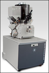
OHSU’s new lab also includes an FEI Helios NanoLab DualBeam, which provides scanning-electron and focused ion beam microscopy.
At the new lab, Gray and other OHSU scientists will be able to visualize cell structures at a high level of detail, enabling them to explore, among other things, how cancer cells function differently as they spread from the site of origin to other parts of the body.
FEI expects that the collaboration will deepen its understanding of the total work flow of electron microscopy in cellular biology and, ultimately, enable it to develop next-generation tools. The focus of this work will be on combining electron microscopy with other imaging techniques, such as light and fluorescence microscopy.
The Living Lab will be part of the OHSU Center for Spatial Systems Biomedicine, which combines physics, biomedical engineering, chemistry and biology to study cancer and other diseases.
For more information, visit: www.ohsu.edu
/Buyers_Guide/FEI/c4773
Published: September 2011
Glossary
- scanning electron microscopy
- Scanning electron microscopy (SEM) is an advanced imaging technique used in microscopy to obtain high-resolution, three-dimensional images of the surfaces of solid specimens. SEM achieves this by using a focused beam of electrons to scan the specimen's surface, resulting in detailed images with magnifications ranging from about 10x to 100,000x or higher.
Key features and principles of scanning electron microscopy include:
Electron beam: SEM uses an electron beam instead of visible light for...
AIDSAmericasBasic ScienceBiophotonicsBusinesscancerelectron microscopesFEI Co.focused ion beam microscopyHelios NanoLab DualBeamImagingJoe GrayLaboratoriesLiving Lab for Cell BiologyMicroscopyOHSUOHSU Center for Spatial Systems BiomedicineOregonOregon Health & Science UniversityResearch & Technologyscanning electron microscopyTitan Kriostransmission electron microscopy