Dr. Barbara Stam, Dr. Gerhard Holst and Ursula Buczek
Changes in bruise color allow doctors to draw conclusions about when an injury occurred.
To determine physical abuse of a child, several warning signs must be considered: Is the child scared? Are bruises present, and if so, where? What do the parents say happened to the child? Although a physician can pick up on most of these signals, a conviction based on this information alone can be difficult.
One reason is the uncertainty as to exactly when the injury happened – a bruise or hematoma bears no time stamp. If it were possible to determine the precise time that the child got hurt, even days later, physicians and courts would have an effective weapon against child abusers.
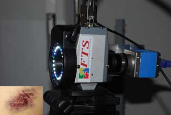
Dr. Barbara Stam of the Academic Medical Center of the University of Amsterdam and colleagues have developed a system that uses the color of a bruise (inset) to determine its age. Here, the ring of LED lights they developed for the system.
Images courtesy of PCO.
A reliable method of calculating that specific moment may be available in the near future, thanks largely to the work of Dr. Barbara Stam (co-author of this article), a medical physicist at the Academic Medical Center of the University of Amsterdam. In her doctoral thesis, Stam laid the groundwork for reliably determining the age of hematomas based on their color. This has been tried in the past, but dating injuries by using color tables is an imprecise method that cannot be used in court. Even technical processes have brought no advances up to now.
Stam built on work carried out by a colleague, Lise Lyngsnes Randeberg of the Norwegian University of Science and Technology in Trondheim, who developed a model linking the color of the hematoma to its age, enabling calculation of when the injury originally occurred. Stam, meanwhile, has succeeded in developing a method based on this model that doctors should be able to use in several years without undue expense or effort.
In the case of a hematoma, the clock starts ticking as soon as the the skin receives the blow. Blood from damaged vessels spreads throughout the surrounding tissue, creating a dark reddish-blue mark, which expands slightly over the next 24 hours. This is caused by hemoglobin, the red blood pigment. At the same time, a larger yellow area of bilirubin forms around the mark, a result of enzymatic conversion of the hemoglobin. The bilirubin is eventually cleared by the lymphatic system, one of whose tasks as part of the immune system is to filter waste products from the body.
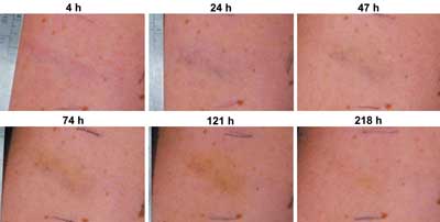
Changing colors in a bruise: At 4 hours (h), only redness is visible. At 24 h, the predominant color is blue, whereas a day later, yellow is also apparent. After 74 h, yellow is the predominant color, but blue is still visible. After 121 h, only yellow is present. At 218 h, a small amount of yellow is still visible. At 264 h, no yellowness is still visible (not shown). The bruise, resulting from a strike with a small cane, is located on the upper arm of a 27-year-old woman.
Key to establishing a bruise’s age is the fact that the two substances increase and disappear at different rates, resulting in an inhomogeneous distribution of color beneath the skin. The dark hemoglobin mark usually reaches its maximum size after just a day; after that, it begins to shrink as it is converted into bilirubin. On average, this mark completely disappears after about a week. In contrast, the yellow area continues to expand for an average period of four days before slowly beginning to disappear.
The presence of the bilirubin is a delicate balance between formation of bilirubin from the breakdown of hemoglobin and clearance of bilirubin into the lymphatic system. This mark also disappears after 10 days. Information about the age of a hematoma is obtained from the ratios between the sizes of the two marks, rather than from the absolute quantity of both, and in particular from the change in these ratios over time.
Bruises, up close and personal
Stam’s apparatus can automatically measure this color distribution. She positions a pixelfly qe camera system from PCO of Germany above the damaged tissue. A liquid crystal tunable filter that allows only certain wavelengths of visible light to pass through it is mounted in front of the camera. The filter can be opened and closed in 2-nm steps within the visible light spectrum between 440 and 700 nm. In the course of a full measurement, 131 images are recorded, each of which is for only a narrow 2-nm band of wavelength. The final result is a 3-D data record that shows the light intensities for each point on the surface of the skin, and for each of the wavelength windows.
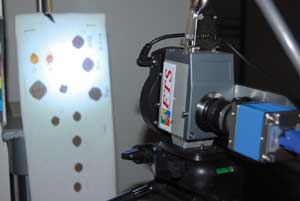
The system uses a tunable acoustic filter (the gray device with the FTS sticker) and a CCD camera from PCO (the blue device). Everything is mounted on a camera stand. Here, the system is shown measuring bloodstains, an alternative use developed by Stam and colleagues.
The recordings measure the intensity of light reflected by the skin. Stam illuminates the hematoma with a ring of white, blue and cyan-colored LEDs, and the recording apparatus adjusts sensitivity fluctuations. One portion of the light is absorbed by the tissue, while another portion is reflected; the reflected portion changes according to whether more hemoglobin or more bilirubin is present in the tissue. Some wavelengths yield especially striking results: Hemoglobin, for example, absorbs mainly at wavelengths of 430, 540 and 575 nm, while bilirubin absorbs at 470 nm. Making the distinction, however, is not easy. The camera’s sensitivity ensures the quality of the recordings.
The entire measurement takes roughly 90 seconds, the limiting factor being the slowness of the electronic filters currently available – the camera alone would be much faster. Stam therefore takes a different approach to speed up the process: If the initial results are confirmed, and just four or five wavelengths of visible light are actually enough to obtain clear “fingerprints” of hemoglobin and bilirubin, fewer recordings are taken, reducing measuring time to several seconds.
Small, portable, cheap
The measuring system is mounted on a small trolley, but the camera, filter and laptop could also fit into a bag; the aim is to make the measuring device cheap enough so that every hospital can afford one. Stam expects that four more years of research will be needed before the measuring technology is sufficiently developed for hospital use.
The process would also be a great help in forensic medicine to uncover cases of child abuse. The results Stam has achieved on hematomas on voluntary test subjects have been very promising. With a bruise that is 24 hours old, Stam can accurately determine the time of the injury to within half an hour. The uncertainty period for 5-day-old bruises that already have turned a clear yellow is still just under 16 hours.
Meet the authors
Dr. Barbara Stam studied medical natural sciences and achieved her master’s degree in medical physics in 2006 at the Free University in Amsterdam. Dr. Gerhard Holst is head of research at PCO in Germany. Ursula Buczek is a public relations consultant and account manager at Storymaker PR in Tübingen, Germany; email: [email protected].