
Chemical Treatment Makes 3-D Cell Cultures Clearer
A chemical treatment could lead to better imaging of cells in 3-D cultures.
A team at Brown University made the finding while studying how neural tissues grow from stem cells. The researchers created sphere-shaped cell cultures to allow the cells to develop more naturally. The thickness of the cultures hampered transparency of the cells, however, making proper imaging difficult.
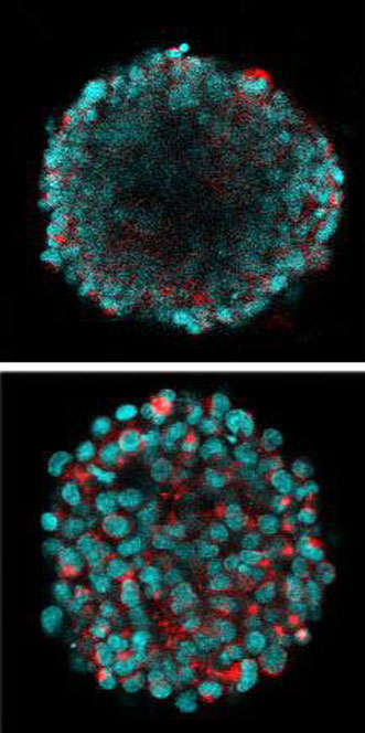
Without a clearing treatment, a 3-D tissue culture, top, does not allow researchers to see deep inside. ClearT2, one of three new chemical treatments evaluated in recent tests, makes tissues transparent, bottom, so that researchers can study cell interactions beneath the surface. Courtesy of the Hoffman-Kim Lab/Brown University.
“As I was imaging these tissues I was only able to get the outer layer or two of cells, and that wasn’t a very good representation of what was going on inside of the sphere,” said Molly Boutin, a graduate student in biomedical engineering.
Rather than dividing the tissues to allow better imaging, the researchers compared three chemical treatments designed to make tissue transparent: ClearT2, SeeDB and Scale.
The ClearT2 technique was found to be the most efficient, as it allowed the researchers to see fluorescing cells at all depths of focus without changing the size of the tissue. Maintaining the size of the tissue culture allows the researchers to determine its physical dimensions of growth.
Scaffold-free, self-assembled adult hippocampal neural stem cells that were 100 µm in diameter could be optically cleared and imaged using ClearT2 while retaining their size, the researchers wrote in the study. With the Scale protocol, the sphere-shaped cell cultures became substantially larger; SeeDB made them smaller and didn't improve clarity as much.
The work was funded by the National Science Foundation, the National Institutes of Health and Brown’s Institute for Brain Science. The research was published in Tissue Engineering Part C: Methods (doi: 10.1089/ten.TEC.2014.0296).
For more information, visit www.brown.edu.
Published: September 2014