
Photoacoustic Imaging Technique Measures Periodontal Health
Using squid ink as a contrast agent for a photoacoustic imaging technique, researchers have developed a dental imaging method for assessing gum health that is noninvasive, comprehensive and accurate. The photoacoustic method can image the entire pocket depth around the teeth at once, without the poking and prodding that typically accompanies the use of a periodontal probe, considered the “gold standard” tool for periodontal examinations. In addition to being invasive and even painful for the patient, the periodontal probe allows only one measurement to be taken at a time.
For the photoacoustic method, researchers at University of California, San Diego, use a mouth rinse made from commercially available food-grade squid ink combined with water and cornstarch as the contrast agent. Squid ink naturally contains melanin nanoparticles that absorb light. Rinsing the mouth with the squid ink mix causes the melanin nanoparticles to become trapped in the pockets between the teeth and gums.
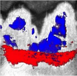
Photoacoustic/ultrasound image after squid ink oral rinse treatment. Ultrasound image of the teeth is in black and white. The photoacoustic signal from the squid ink contrast agent in the pocket depth is in red, and signals from stains on the teeth are in blue. Courtesy of Jokerst Bioimaging Lab at UC San Diego.
When researchers shine a laser light onto the area, the squid ink heats up and quickly swells, creating pressure differences in the gum pockets that can be detected using ultrasound. This method enables researchers to create a full map of the pocket depth around each tooth.
Researchers tested the photoacoustic imaging method in a pig model containing a mix of shallow and deep pockets in the gums.
Thirty-nine porcine teeth (12 teeth with artificially deeper pockets) were treated with the contrast agent, and the probing depths were measured with photoacoustic imaging and with a Williams periodontal probe. There were statistically significant differences between the two measurement approaches for distal, lingual and buccal sites, but not mesial. The photoacoustic imaging approach offered 0.01-millimeter precision and could cover the entire pocket, as opposed to the probe-based approach.
While results using the photoacoustic imaging method closely matched measurements taken using a periodontal probe, they were also consistent across multiple tests. Measurements with the periodontal probe varied significantly from one test to another.
“Using the periodontal probe is like examining a dark room with just a flashlight and you can only see one area at a time. With our method, it's like flipping on all the light switches so you can see the entire room all at once,” professor Jesse Jokerst said.
The team believes that their innovation could be a significant improvement over the conventional method for evaluating gum health.
“It’s remarkable how reproducible this technique is compared to the gold standard,” Jokerst said.
Moving forward, the team will be collaborating with dentists and testing their method in humans. Future work also includes minimizing the salty, somewhat bitter taste of the squid ink oral rinse and replacing laser lights with inexpensive, more portable light systems like LEDs. The team’s ultimate goal is to create a mouthpiece that uses this technology to measure periodontal health.
The research was published in the Journal of Dental Research (doi: 10.1177/0022034517729820).
A video Q&A with professor Jokerst, on a noninvasive, photoacoustic imaging method for conducting an accurate, comprehensive periodontal exam. Courtesy of UC San Diego Jacobs School of Engineering.
Published: September 2017