Ya Sha Yi, University of Michigan, and Bo Liu, Tongji University
Combining the principles of integrated nanophotonics with traditional scintillator materials will greatly improve the development of radiation detection. Strong evidence exists that integrated nanophotonics innovations will have an evolutionary – and revolutionary – impact on this field, especially for next-generation high-performance medical imaging instruments.
Scintillators have been critical to biomedical imaging, including brain imaging, as well as to high-energy particle detection and explosives detection for national security. Integrated nanophotonics will significantly boost the light extraction efficiency and directional light emission control of medical-imaging scintillator materials and devices to enable lower-dose, higher-timing/energy/position/resolution medical imaging applications.
The earliest scintillator, CaWO4, was discovered in 1896, which was followed by Röntgen’s discovery of the x-ray. In the 1940s, the modern radiation-detection system emerged, thanks to the development of the photomultiplier tube and the discovery of NaI:Tl, which is the most typical scintillator.
Scintillators are materials that absorb incident particles (such as electrons, protons, neutrons, α-particles) or high-energy photons (such as x-rays and γ-rays). They subsequently convert the deposition energy efficiently into a large number of photons in the visible or UV range, which can be directly detected by photomultipliers, photodiodes or CCDs. In this way, the energy, position and time of incident radiation can be determined efficiently. Scintillators play an important role in radiation-detection systems with various applications in medical imaging, high-energy particle experiments and national security.
The scintillation process involves spontaneous emission under the excitation of ionizing radiation. The physical processes involved in scintillation can be divided into three stages. The first is the ionization event, which creates inner shell holes and primary electrons via photoabsorption interactions, Compton scattering and electron-positron pair formation. The second stage is the thermalization of hot electrons and holes, until the energy becomes less than the ionization threshold. The carriers diffuse in the lattice and are subsequently trapped on defects and impurities, which can lead to excitation of the luminescence centers. The third and final stage is the luminescent process, in which the excited luminescent center returns to ground state by emitting a photon or nonradiative transition.
In general, an ideal scintillator has the following properties: high light yield, short decay time, high density, no afterglow, good spectral match to photodetectors, good chemical stability and mechanical strength, and low cost. Light yield is defined as the number of photoelectrons generated by radiation with a unit of energy. The measured light yield also must take into account the light-extraction efficiency and quantum efficiency of the photodetector.
High light yield of a scintillator is vital to improving sensitivity, spatial resolution and energy resolution of detection systems. For one example, with improved sensitivity, we can apply a lower radiation dose during medical imaging procedures (such as x-ray CT), resulting in a corresponding decrease of cancer risk due to radiation exposure. Short decay time is critical for improving time resolution for particle detection. This is particularly important in the application of positron emission tomography (PET) scans with the coincidence time technique, in which a shorter time window is applied, leading to a reduced background of random events and, thus, better-quality imaging.
Generally, a high-density scintillator composed of elements with large atomic numbers has strong stopping power for the incident radiation, which makes the detector more compact. A compact detector not only can largely reduce cost, but also can improve the spatial resolution in imaging systems.
However, an ideal scintillator – combining all advantageous properties – does not exist in practice, so trade-offs are necessary. For a given detection system, the most suitable combination of properties from available scintillators depends on user requirements as a function of
priority.1
Development challenges
In recent decades, the need for medical imaging systems and high-energy particle experiments has promoted ongoing development of scintillators. New scintillators with high light yield and fast decay times have been proposed and have found many practical applications. For example, the LYSO ([Lu,Y]2SiO5:Ce) scintillator is a promising candidate in PET systems, and the PbWO4 (PWO) scintillator has been used in the Compact Muon Solenoid of the Large Hadron Collider at CERN, the European Organization for Nuclear Research.
Light yield is the most important requirement in most applications. Exploring scintillators with a much higher light yield than usual has attracted much attention in the field. For example, in recent years, researchers proposed LuI3:Ce with light yield of 76,000 phe/MeV, the highest value among inorganic scintillators. Although much effort has been devoted to exploring new scintillators with high light yield based on crystal structure and elemental constituent, it is increasingly difficult to find new materials with unique properties.
Most inorganic scintillators have a high refractive index (n ≈ 1.8 to 2.2), which is much higher than the ambient medium. The light created inside the scintillator could be totally reflected at the crystal-air interface if the incident angle is larger than the critical angle. These photons cannot reach the detectors but emit at the side face or are absorbed during multireflection.

In practical applications, only photons that can enter detectors are useful. Therefore, we must define effective light yield as in Equation 1, where LYabs is absolute light yield, defined as photons created by radiation with unit energy; ηex is the efficiency of light extraction, indicating the ratio of photons that can emit out of the scintillator. LYeff is effective light yield.
The total internal reflection is the main reason for low extraction efficiency. According to Snell’s law, light can emit from a scintillator when the incident angle is smaller than the critical angle. If we neglect Fresnel reflection, considering an infinite plane scintillator-air interface, the extraction efficiency from one side face can be determined as in Equation 2.
Assuming that the refractive index of the scintillator is 2, extraction efficiency is about 6.7% as calculated in Equation 2. In some cases, extraction efficiency is enhanced by an optical coupling with grease between the scintillator surface and the photodetector window. It is possible to further improve light extraction because the coupling cannot totally eliminate the difference in refractive indices. In some specific cases, the optical coupling cannot be applied, and thus efficient light extraction becomes extremely important.
For high-index scintillators with a planar surface, assuming a point-like source located a short distance below the scintillator-air interface, the far-field emission exhibits a Lambertian pattern following a cosine dependence on the angle. Emission intensity reaches maximum at the normal direction and decays with the increasing angle. This emission profile is disadvantageous to the detection system because, in general, only a fraction of the total emitted light enters the detector, as illustrated in Figure 1. It would be better to design the directionality of emission according to the layout of systems and aimed at maximum photon collection.
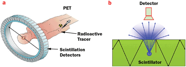
Figure 1. (a) Scintillator devices are the core part of biomedical imaging instruments, including those used for brain imaging. (b) This general schematic sketch of the layout of a scintillation detection system indicates the propagation of emission inside and outside the scintillator. Photo courtesy of The Optical Society.
Integrated nanophotonics on scintillator detectors
Integrated nanophotonics deals with the interaction between light and material on the nanoscale. This interaction leads to strong control of the propagation of electromagnetic waves and the control of optical mode density distribution, which essentially influences the rate of spontaneous radiation. The types of nanostructured materials may include photonic crystals, surface plasmon polaritons or optical microcavities. The structure scale is on the order of wavelength or subwavelength. Photonic crystals are nanostructured materials in which alternating dielectric components with higher and lower refractive indices create a periodic structure on the order of the wavelength of light. Metallic nanoparticles exhibit unique optical properties due to the excitation of surface plasmon polaritons, which usually belong to the subwavelength scale.
Next we will explore some examples that illustrate scintillator principles and applications.
A nature-inspired photonic structure
A nature-inspired moth-eye structure (comprising an array of circular protuberances such as those on a facet in a moth’s eye) provides a unique method to achieve broadband antireflection. This bioinspired structure can be used to enhance light extraction of the scintillator due to periodicity. We have reported that an improved moth-eye structure – adding roughness on the sidewall of the original pyramid shape – can achieve extraction enhancement of up to 2.7 when periodicity is 400 nm.2 The structure was fabricated via the process illustrated in Figure 2a. The SiO2 nanoparticles in the mask layer were coated on the high-index Si3N4 layer, which was previously deposited on the Lu2SiO5:Ce (LSO) scintillator thin film at a thickness of 500 nm. After reactive ion etching and removal of the remaining SiO2 nanoparticles, the scanning electron microscope (SEM) image shown in Figure 2b exhibits the improved, bioinspired moth-eye structure. The finite-difference time-domain (FDTD) simulations in Figure 2c show the optimal structure parameter and the enhancement. The experimental characteristic of the emission spectra, with reference and structured samples under x-ray excitation, exhibits significant enhancement with the broadband, shown in Figure 2d. The method of using a mask is superior to conventional e-beam lithography because of its ability to fabricate samples with large-scale area, which is important to certain practical applications.
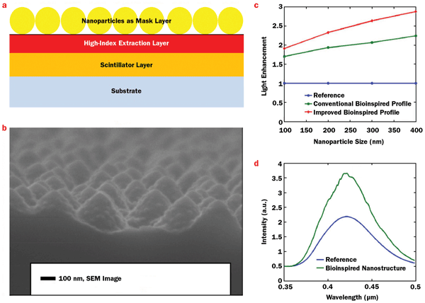
Figure 2. (a) The self-assembly of SiO2 nanoparticles on top of high-index light-extraction layer Si3N4, which is deposited on a Lu2SiO5:Ce (LSO) thin film. (b) This SEM image of the improved, bioinspired moth-eye nanostructures with a certain degree of roughness on the sidewall shows interesting nano-on-nano features. (c) Finite-difference time-domain simulation results, which show light-output enhancement for both bioinspired light-extraction structures, while the improved bioinspired moth-eye structures show more light-output enhancement. (d) A comparison of light output between two LSO thin films from the excitation of an x-ray mammographic unit with a molybdenum target. The blue curve is light output from the referenced sample, and the green curve is light output from sample with the improved, bioinspired moth-eye structure. Figure courtesy of The Optical Society.
These moth-eye-like structures are widely used as antireflection coatings in applications where the pyramid shape plays the role of gradual-refractive-index layer, and therefore the Fresnel reflection can be significantly eliminated. However, the periodic structure is the most important factor for light extraction because it provides the reciprocal lattice vector, adding to the in-plane vector of guided modes. The individual pyramid shape may optimize extraction efficiency.
Photonic crystals with monolayer periodic nanospheres
We have demonstrated the significant enhancement of light extraction for bismuth germinate (BGO)3 and LYSO4 scintillators in a broad emission spectrum using a monolayer of a periodic array of polystyrene (PS) nanospheres. Schematic diagrams and top-view SEM images appear in Figures 3a-c. The SEM image of the self-assembled monolayer of 500-nm-diameter PS nanospheres on the BGO scintillator surface exhibits a hexagonal close-packed structure with two directions: Γ–M and Γ–K.
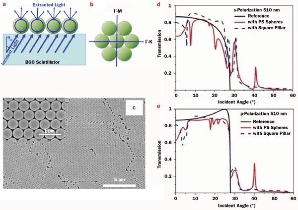
Figure 3. (a) A schematic of light extraction for a bismuth germinate (BGO) scintillator with a monolayer of polystyrene (PS) nanospheres. (b) Sketch of the two different directions of the hexagonal close-packed structure. (c) Top-view SEM images of the surface of BGO covered with PS nanospheres (d = 500 nm) with two different resolutions. Numerical simulations of transmission spectra with a wavelength of 510 nm for plain BGO (black solid lines); BGO covered with a monolayer of PS nanospheres with a diameter of 500 nm (red solid lines); and BGO covered with an array of square pillars with the same structures as the monolayer of PS spheres (blue dashed lines). The simulations with s- and p-polarizations are shown in (d) and (e), respectively. Photo courtesy of John Wiley & Sons.
To illustrate the effect of the nanosphere monolayer on light extraction, numerical simulations for transmission at the maximum emission wavelength of 510 nm are shown in Figures 3d and 3e for s- and p-polarization, respectively. For the plain-surface BGO, transmission drops to zero beyond the critical angle of 27.7°. After being covered with the nanosphere monolayer, it is fortunate to find that two extra transmission peaks appear at about 30° and 40° for both polarizations. This implies that incident light beyond the critical angle can be partly extracted, leading to enhanced extraction efficiency.
The photoluminescence spectra of BGO in Figures 4a and 4b clearly show enhancements after covering with a monolayer of PS spheres; they also are strongly dependent on wavelength and emergence angles. The wavelength- and angle-integrated enhancement reaches 72 percent. To obtain optimal extraction efficiency, the diameter must be small enough to ensure that only low-order waveguide modes are excited, and at the same time, it should be large enough to ensure that diffraction can take place effectively.
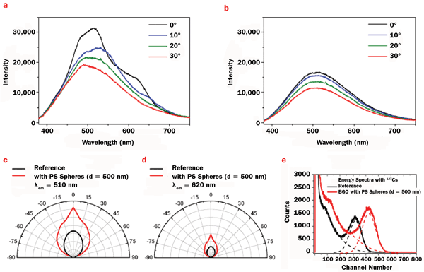
Figure 4. The emission spectra of BGO covered with a monolayer of PS nanospheres (a) and plain BGO (b) with different emergence angles. Angular profiles of emission at 510 nm (c) and 620 nm (d) for plain BGO (black lines) and BGO with nanospheres (red lines). The light output (e) under the excitation of γ-ray for plain BGO (black lines) and BGO with nanospheres (red lines). Gaussian fittings, shown with dashed lines, are used to analyze the energy resolution. Photo courtesy of John Wiley & Sons.
Experimental and simulated results suggest that the optimal diameter is close to the wavelength involved. The angular profile of emission intensities at 510 and 620 nm shown in Figures 4c and 4d are significantly increased in the whole emergence angle, with strongest enhancement at normal direction, making the angular profile deviate from the Lambertian pattern. The directivity of emission is dependent upon the diffraction of the structure of periodic array. This angle-dependent enhancement would be advantageous to practical applications, because only photons emitted in the direction of the detector window can contribute to the detection.
From the viewpoint of application, it is therefore necessary to design the scintillators’ structures carefully, according to the layout of a system for a certain purpose of radiation detection. The energy-spectrum measurements for γ-rays from a cesium-137 source are shown in
Figure 4e. The energy spectrum reflects the wavelength and angle integration of scintillation photons. For the photopeak, the channel number (light output) of the structured BGO is larger than that of the reference sample by 34 percent. It should be noted that the structured BGO is covered with PS nanospheres in half of the area. Therefore a corrected enhancement of light output is expected to be about 68 percent. It is interesting to observe that energy resolution for the photopeak is improved from 33.6 percent (in a reference sample) to 30.3 percent (in a sample with PS nanospheres), deduced from experimental data using Gaussian fitting.
The obvious advantage of the self-assembled method lies in the ability to fabricate samples with a large-scale area, which is important to the application of scintillation detection. Furthermore, using self-assembled patterns as templates, more complicated photonic structures can be fabricated, which expands compatibility and the ability to control the propagation of light. This work is still in progress.
Meet the authors
Ya Sha Yi is an associate professor and director at the Integrated Nano Optoelectronics Laboratory at the University of Michigan, Dearborn, and an associate professor at the Energy Institute at the University of Michigan, Ann Arbor; email: [email protected]. Bo Liu is a professor at the School of Physical Science and Engineering at Tongji University in Shanghai; email: [email protected].
References
1. M.J. Weber (2002). Inorganic scintillators: today and tomorrow. J Lumin, Vol. 100, pp. 35-45.
2. P. Pignalosa et al (2012). Giant light extraction enhancement of medical imaging scintillation materials using biologically inspired integrated nanostructures. Opt Lett, Vol. 37, pp. 2808-2810.
3. Z. Zhu et al (2014). Broadband light output enhancement for scintillator using whispering-gallery modes in nanospheres. Phys Status Solidi A, Vol. 211, pp. 1583-1588.
4. Z. Zhu et al (2013). Improved light extraction efficiency of cerium-doped lutetium-yttrium oxyorthosilicate scintillator by monolayers of periodic arrays of polystyrene spheres. Appl Phys Lett, Vol. 102, 071909.