Compiled by BioPhotonics staff
A three-dimensional cell imaging method has been developed to study the
complex spatial-temporal dynamics of protein transport, a challenging feat in the
field of cell biology.
Because of cell-to-cell variations in thickness and the temporal
properties of protein transport, scientists have faced many technical challenges
trying to image such dynamic processes in the cell and in 3-D. Previous techniques
were slow and suffered from poor Z-localization and 3-D tracking capability, say
researchers at the University of Texas Southwestern Medical Center at Dallas and
the University of Texas at Dallas in Richardson.
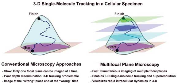
Illustrated are the strengths of multifocal plane microscopy over conventional microscopy techniques. Courtesy of S. Ram, UT Southwestern Medical Center.
To overcome the obstacles, the scientists used a combination of
multifocal plane microscopy and nanodot labeling technology. They labeled single
molecules in live cells and tracked their movement and interaction with other molecules
in a thick cell sample over an extended period.
Initially, the technique was developed by the researchers to track
the movement of therapeutic antibodies engineered in their lab. Although current
microscopy technologies limit scientists to image only a single focal plane at a
time, the scientists wanted to simultaneously image a sample across multiple planes.
Their findings, which were funded by the National Institutes of
Health and the National Multiple Sclerosis Society, were reported at the 55th Biophysical
Society Annual Meeting in Baltimore.