
Build Versus Buy: Considerations in SD-OCT System Design
The spectrometer used in an OCT system, whether custom-built or preconfigured, has a direct impact on both the quality of images acquired and the ease and speed of system design.
CICELY RATHMELL, WASATCH PHOTONICS
Many researchers and product developers using spectral-domain optical coherence tomography (SD-OCT) for biomedical research and clinical treatment choose to build their own system. This requires multiple optical and mechanical components, an understanding of signal and image processing, and the optics and programming expertise needed to bring it all together — as well as a significant investment of time to assemble and calibrate the system. Using a prebuilt, off-the-shelf OCT spectrometer as one of the starting components can speed and simplify this process, reduce risk, and improve the quality of images collected.
This primer will delve into key considerations for the optical designer when building an SD-OCT system, with a focus on how to get the best performance from the OCT spectrometer incorporated into the design — whether the plan is to build the spectrometer or buy one.
OCT gains traction
OCT has gained popularity as an imaging modality due to its ability to provide images with much higher resolution than ultrasound, without the need for direct contact with a specimen or use of a coupling medium. While it was introduced into the mainstream as an ophthalmology technique (Figure 1), OCT is now viewed as a more broadly applicable approach to noninvasive biomedical imaging. It provides surface profiles and information about subsurface structure and uniformity up to a few millimeters in depth, with applications in medical fields such as ophthalmology, dermatology (Figure 2), cardiology, and life sciences research. It also has uses outside the life sciences and medicine, in industrial inspection, and even for the analysis of art and historical artifacts.
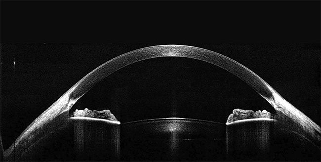
Figure 1. 1300-nm spectral-domain optical coherence tomography (SD-OCT) image depicting the structure of a cornea and anterior segment of the eye. Courtesy of Wasatch Photonics.
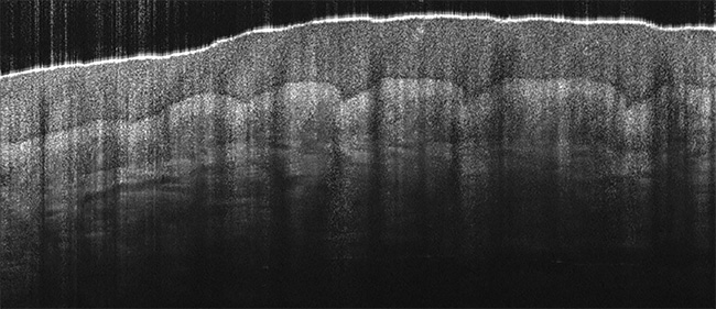
Figure 2. 800-nm spectral-domain optical coherence tomography (SD-OCT) image of a finger, showing the epidermis and dermis layers.
Courtesy of Wasatch Photonics.
The widespread development of both research and commercial OCT systems has made many of the required components more widely available, often in the form of modules that speed and simplify design. This, in turn, has made the technique more accessible and increased the adoption of OCT as a complementary real-time imaging technology in fields such as surgery and dentistry.
For the potential user who is considering the design of a new OCT system, the question becomes whether to build or buy for several of the key elements. Building each individual component allows the system to be customized to meet exact needs but increases the time and risk of the construction, because all subcomponents might not perform as expected or be available as needed. The use of preconfigured modules — optimized specifically for OCT — can improve performance and lower costs in the long run, particularly for commercial system development.
Building a custom OCT system
Both swept-source OCT (SS-OCT) and SD-OCT are widely used for OCT imaging of the eye for the diagnosis of injury and disease, including macular degeneration, retinal damage, glaucoma, and corneal pathologies such as keratoconus. In SS-OCT, the laser is scanned across a particular wavelength band, and a single-element photodetector captures the signal. In SD-OCT, a broadband laser source is used in combination with a multielement detector such as a spectrometer. SS-OCT offers high speed and low roll-off (loss of sensitivity with depth) but can be more expensive due to the high cost of swept-source lasers. SD-OCT yields better resolution at a lower cost, and due to the introduction of better imaging cameras in recent years, high-speed SD-OCT is quite competitive in terms of speed (250 kHz or higher) and roll-off performance for many applications.
In an SD-OCT system, the spectrometer functions as the detector, digitizing the intensity of scattered light at each point on the sample (Figure 3). Due to the interferometric nature of the technique, this spectrum can be transformed into a depth profile. To generate a cross-sectional OCT image, the light source is then swept across the sample with a high-speed scanning mechanism such as a galvanometer or MEMS-based device, capturing a spectrum at each point. Custom software must be written to communicate with both the spectrometer and scanning arm to synchronize data collection with the beam scanning and to process the spectra collected.
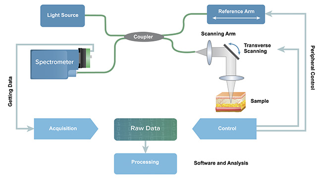
Figure 3. Spectral-domain optical coherence tomography (SD-OCT) system schematic depicting optical design, system control, and data flow. Courtesy of Wasatch Photonics.
The key modules in an SD-OCT system, such as light sources, optical couplers, and reference arms, are produced with advanced manufacturing techniques and can be purchased off-the-shelf. The OCT spectrometer, on the other hand, is somewhat more distinctive because it must be matched to the light source and designed specifically to achieve the desired imaging depth and resolution for research or diagnostic purposes. This complex subunit requires a diffraction grating, custom optical elements, and a specialized camera in precise alignment. It is a particularly challenging module to custom-build effectively, yet its unique requirements — including high spectral resolution, high speed, and polarization independence — are pivotal to an OCT system’s performance.
Keys to the OCT spectrometer
The OCT spectrometer used as the sensor in an SD-OCT system has a considerable effect on the quality of images collected — influencing the imaging depth, resolution, speed of image acquisition, and contrast in the image. Low roll-off (loss of sensitivity with depth) is the key performance parameter for any OCT spectrometer and is determined by the spectrometer design.
Whether one is purchasing or designing an OCT spectrometer, it is important to understand both the optical and camera-related factors that will influence system performance and the ease of system development, each of which is detailed below:
1. High efficiency at all wavelengths
The reflected light in OCT is very low intensity. Therefore, a highly sensitive spectrometer will substantially improve the resulting image quality. And because OCT uses a Fourier transform of the spectrum to generate a depth profile, every wavelength in that spectrum is important. A spectrometer with a highly uniform response as a function of wavelength delivers better image quality, which is essential to the development of reliable diagnostics. This can best be achieved by using holographic transmission gratings, particularly designs patented specifically to maximize efficiency, minimize scatter, and reduce the variation in efficiency across the full range of wavelengths being used.
2. Insensitivity to polarization
OCT is an interferometric technique, and fiber optics are often used for light routing. This results in a system that is highly sensitive to polarization changes. While most of the components in a spectrometer are polarization-insensitive, several of the grating types used in commonly available spectrometers can have a strong polarization dependence and therefore are not suitable for OCT imaging. Volume phase holographic transmission gratings have very low polarization sensitivity, making them an excellent choice for SD-OCT systems.
3. Diffraction-limited optical design
Off-the-shelf optics often fail to deliver high clarity OCT images that resolve fine structures because the optical constraints to obtain good roll-off are so stringent. The spot size of light imaged onto each camera pixel must be small to avoid spreading to neighboring pixels, and this must be true for the broad range of wavelengths used in the spectrometer. By designing and sourcing custom lens sets matched to the rest of the spectrometer design, it is possible to achieve optimized spectrometer performance at all wavelengths, far better than the performance of off-the-shelf lenses (Figure 4).
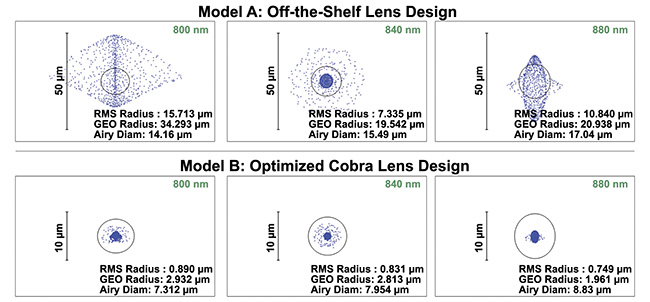
Figure 4. The quality of focus onto an OCT camera varies considerably with wavelength when off-the-shelf lenses are used in an OCT spectrometer design (top). This results in greater roll-off (loss of sensitivity with depth). By comparison, custom lens sets optimized to match the spectrometer design offer far better focusing across wavelength, reducing roll-off considerably (bottom). Courtesy of Wasatch Photonics.
4. A high-speed, high-fidelity camera
While careful optical design can significantly reduce roll-off in an OCT spectrometer, crosstalk between camera pixels may limit the performance that can be achieved, and therefore it is not desirable for clinical applications in which fine structure must be observed. Use of high-sensitivity, low-crosstalk cameras, such as the e2v OctoPlus camera, can minimize this effect; they can also deliver scan rates of 250 to 400 kHz, which is comparable to most commercial SS-OCT speeds.
5. Choice of camera connection
The OCT spectral data that is generated by the camera is transferred through a camera link card or USB cable, depending on the camera model. Camera-link communication allows for faster scan speeds and is a more stable platform for many commercial OCT applications with stringent clinical demands, but it requires the purchase of the requisite camera-link card and a computer in which to install it. Use of USB 3.0 communication, on the other hand, reduces system cost and size, which can be very convenient in mobile medical applications. While USB 3.0 communication does not support the same high rates of data transfer available via camera link connection, increased adoption of USB 3.1 is likely to overcome this limitation. If flexibility is important in your design, look for a supplier who offers both communications options.
6. Software development kits
The cameras used in OCT spectrometers also have various nonmedical uses and come with extensive, complex manuals of commands for control and data acquisition that can be time-consuming to sift through. A considerable amount of time can be saved by choosing an OCT spectrometer that comes with prebuilt software development kits for OCT — a set of streamlined, OCT-specific commands and sample graphical user interfaces. This approach can save days to weeks of software development time and reduce the risk to the development schedule caused by communications challenges.
7. Compliance with regulatory
requirements
Developers of biomedical instruments must also pass the many regulatory requirements that are essential to safe and effective use of the products. These include immunity to external electromagnetic radiation, limitations in emissions of such radiation, and performance across the specified range of environmental conditions: temperature, humidity, and pressure, as well as mechanical shock and vibration. Designing and testing for all these requirements can be time-consuming and expensive. Use of a well-tested spectrometer design that has been widely deployed in the field can reduce this burden.
Purchasing an OCT spectrometer
Choosing to purchase a preconfigured OCT spectrometer instead of building one has many advantages, from improved performance (clearer, more detailed images to greater depth) to considerable time savings in optical design, build, and software development. This allows the system designer to place greater focus on system and application development without compromising image quality.
OCT can be performed at wavelengths ranging from the visible to NIR, depending on the imaging depth required and the desired detail of a sample. The use of a shorter center wavelength, such as 800 nm, increases axial resolution, which is good for high-resolution imaging of the retina. The use of a longer center wavelength, such as 1300 nm, increases the imaging depth in tissue, which is more advantageous for dermatological imaging. It can therefore be beneficial to work with a spectrometer vendor who offers a variety of center wavelengths, bandwidths, and scan speeds. This flexibility makes it much easier to find a spectrometer that meets the desired requirements, because spectrometer center wavelength, bandwidth, and the resulting axial resolution are interrelated (Figure 5). A typical consultation begins with identifying the center wavelength and bandwidth for the depth and resolution needed, before determining the optimal camera, speed, and connection for the application.
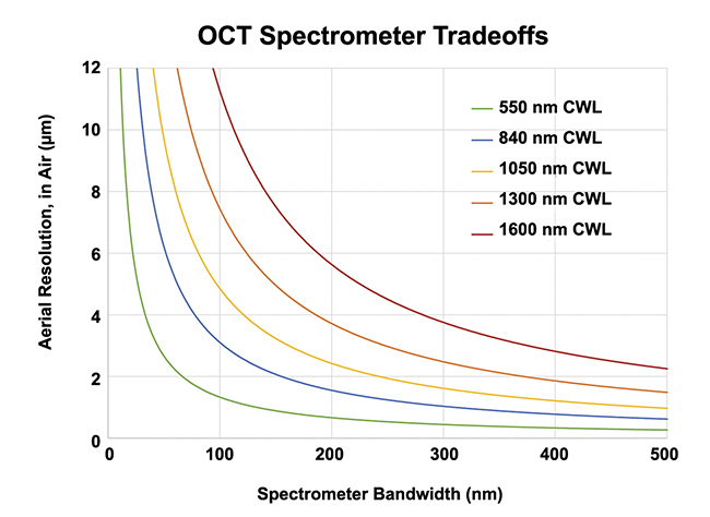
Figure 5. The center wavelength and bandwidth work together to determine the axial resolution of an OCT spectrometer, with longer wavelengths penetrating deeper into tissue and other highly scattering media. Courtesy of Wasatch Photonics.
Streamlined system design
Designing an OCT system for commercial use — whether as a standalone ophthalmologic instrument or complementary imaging for surgical guidance — could warrant additional size and integration restrictions. In this case, it can be worthwhile to source one of the more streamlined, ruggedized OCT spectrometers on the market designed specifically for OEMs.
The best among these are less than half the size and weight of a research-grade OCT spectrometer, weighing <1 kg, yet offer comparable performance in terms of roll-off and speed (Figure 6). This design is robust enough to meet the stringent requirements for thermal and mechanical performance, which are also required in medical devices and mobile surgical equipment. By matching a compact OCT spectrometer in wavelength to an equally compact and cost-effective superluminescent diode light source module, it is possible to achieve economical OCT imaging in volume.
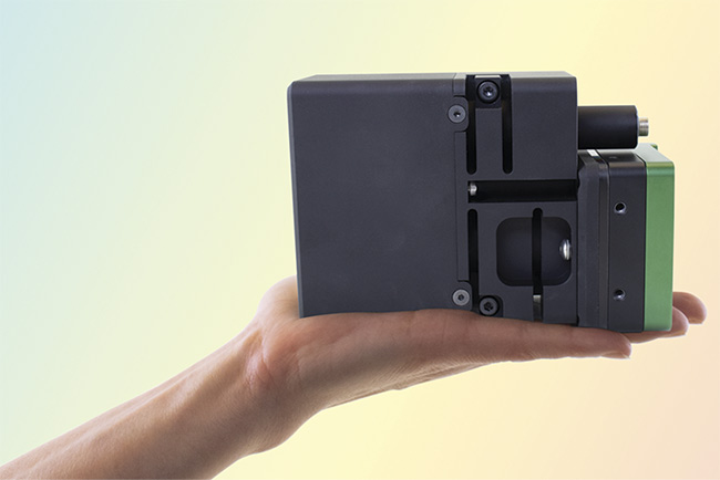
Figure 6. Today’s compact OCT spectrometers are small enough to fit in the palm of a hand, making them suitable for size-constrained
applications in the clinic or operating theater. Courtesy of Wasatch Photonics.
Perhaps the greatest advantage of purchasing a preconfigured OCT spectrometer, however, is the elimination of uncertainty and guarantee of quality. As a module, it is specified to meet a defined set of performance parameters, from the first unit to the hundredth. It is backed by the manufacturer’s quality system and validation testing. This consistency of build translates into more consistent and high-quality image acquisition, thereby increasing the accuracy of interpolated measurements and image analysis.
A pre-built OCT spectrometer allows the researcher, system designer, and/or manufacturing floor to replace the complexity of specifying, controlling, and building a high-performance spectrometer with a single, purpose-ready component that simply functions. This, in turn, can reduce the cost, risk, and time needed to develop and build an OCT system, whether in the lab or in volume.
Due to the introduction of many high-quality, ready-made subcomponents, building an SD-OCT system is easier to accomplish than it used to be and its popularity continues to grow among non-ophthalmology applications. Whether you have a new application or an existing system, purchasing a preconfigured spectrometer is worth considering to accelerate system design and improve image quality. And when system developers can focus on the scientific and/or clinical detail that results — rather than the minutiae of individual component design — medical research and patient outcomes benefit most.
Meet the author
Cicely Rathmell is vice president of marketing at Wasatch Photonics. She has a Master of Science in laser-based atompic spectroscopy from McMaster University. Her 25-year photonics career spans research, product management, sales, and marketing of both components and systems; email: [email protected].
/Buyers_Guide/Wasatch_Photonics_Inc/c15957
Published: September 2023
Glossary
- spectrometer
- A kind of spectrograph in which some form of detector, other than a photographic film, is used to measure the distribution of radiation in a particular wavelength region.
- galvanometer
- An instrument for detecting or measuring small electric currents.
- fourier transform
- Any of the various methods of decomposing a signal into a set of coefficients of orthogonal waveforms (trigonometric functions).
- polarization
- Polarization refers to the orientation of oscillations in a transverse wave, such as light waves, radio waves, or other electromagnetic waves. In simpler terms, it describes the direction in which the electric field vector of a wave vibrates. Understanding polarization is important in various fields, including optics, telecommunications, and physics.
Key points about polarization:
Transverse waves: Polarization is a concept associated with transverse waves, where the oscillations occur...
- ophthalmology
- Ophthalmology is a branch of medicine that focuses on the anatomy, physiology, and diseases of the eyes and visual system. Ophthalmologists are medical doctors who specialize in the diagnosis, treatment, and prevention of eye disorders and diseases. They are trained to provide comprehensive eye care, including medical, surgical, and optical interventions.
Key areas within ophthalmology include:
General eye care: Ophthalmologists perform routine eye examinations to assess visual acuity,...
- optical coherence tomography
- Optical coherence tomography (OCT) is a non-invasive imaging technique used in medical and scientific fields to capture high-resolution, cross-sectional images of biological tissues. It provides detailed, real-time, and three-dimensional visualization of tissue structures at the micrometer scale. OCT is particularly valuable in ophthalmology, cardiology, dermatology, and various other medical specialties.
Here are the key features and components of optical coherence tomography:
Principle of...
SpectrometeroctSD-OCTSS-OCTgalvanometerMEMSOEMFourier transformpolarizationophthalmologydermatologyFeaturesoptical coherence tomography