Due to its nondestructive performance, this technology is well positioned for the continuous monitoring of cell health and differentiation and the quality assurance of cell therapies.
KERSTI ALM AND LISA BODILY, PHASE HOLOGRAPHIC IMAGING
Regenerative medicine is defined as the process of replacing, engineering, or regenerating human or animal cells, tissues, or organs to restore or establish their normal function. Researchers have developed techniques and devised innovative treatments using methods that are suited for small-scale cell production in standard laboratories. For example, in an ongoing Swedish-British study, eight patients with Parkinson’s disease will receive stem cell treatment. However, before any cell treatments can become available to the general population, production must be scaled up, streamlined, and safe. To scale up the process, larger batches of cells must be produced, and the batches must be standardized and monitored to invariably deliver the expected outcome safely. A nondestructive technology such as quantitative phase imaging (QPI) is capable of meeting this need.
Cells and cell culturing are the basis for all biomedical development. Unfortunately, cells are almost invisible by nature, causing direct analysis during culturing to be challenging. Over time, many different methods have been developed to visualize cells, including several types of light microscopy, electron microscopy, and atomic force microscopy. But these techniques are all, to some degree, destructive of the cells, and therefore inhibit undisturbed live cell imaging. Either the light intensity causes phototoxicity, or the samples must be fixed and coated with a reflective metal before imaging. QPI, on the other hand, is one of the very few methods that allows for cell monitoring, imaging, and quantification without disturbing or harming the cells in any way. QPI has been used to monitor the way in which individual cells change over time, and also how entire populations of cells are affected, for example, by drug treatments or environmental changes.
Cell-friendly imaging
Microscopic techniques normally entail focusing intense light on cells followed by the image capture. This focused, intense light causes phototoxicity or damage to the cells’ functioning or structure. In addition, stains or labels are often necessary to enable the cells to be sufficiently visible for imaging, but this can also have a negative effect on the cells that are under study.
QPI, on the other hand, does not require any light to be focused on the cells. Also, QPI does not involve using destructive labels or stains to image and quantify the cells. Instead, QPI exploits the very slight delay of light as it passes through cells. After capture, a computer algorithm converts the slight delay, also known as a phase shift, into a determination of cell thickness. The software can then create topographical maps of the cells1. In other words, all QPI images are reconstructed and are not actual images (such as those taken with a camera) (Figure 1). In addition, pseudo-colors are often added to the resulting images to indicate the cell thickness calculated by the algorithms.
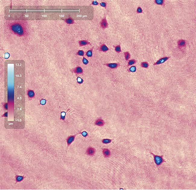
Figure 1. A reconstructed holographic image captured using HoloMonitor M4 from Phase Holographic Imaging. The colors correspond to cell thickness, as shown by the color bar. The ruler indicates cell size. Courtesy of Phase Holographic Imaging.
Since it is cell-friendly, QPI is well suited for the monitoring and quantification of growing and multiplying living cells. Furthermore, the ongoing long-term monitoring and real-time quality assurance of cells is possible by using an imaging device that is compatible with a relevant physical growth environment for the cells, such as a cell culture incubator.
There are many types of QPI, including holographic tomography, digital holographic microscopy (DHM), and spatial light interference microscopy. Some microscope setups will allow for the detailed study of individual cells, while others aim to study cell populations.
Setting up a DHM experiment
The process of setting up a DHM experiment in a laboratory is straightforward: Seed and treat cells in a suitable culture vessel, then place the cells on the microscope stage. Set the capturing positions, imaging frequency, and experiment length, then start the automatic image capture and wait for the experiment to conclude.
The images that DHM captures are not images of the cells. Instead, the light that passes through the cells is merged with a reference light, and the resulting interference pattern — the hologram (Figure 2) — is captured on a sensor. The hologram contains information regarding the amount of light that was delayed as it passed through the cells. The denser the cell, the more the light was delayed. A computer algorithm converts the holograms to topographic cell maps (Figure 2), which allows the cells to be viewed in 3D.
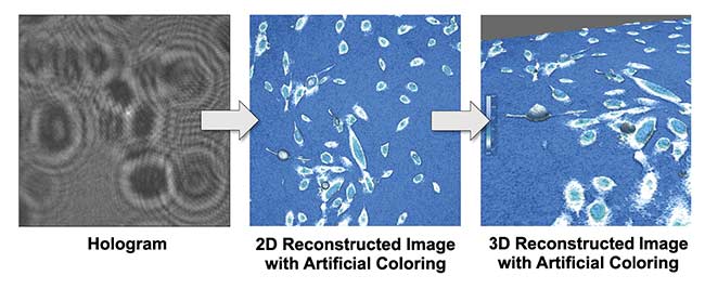
Figure 2. A hologram is the basis for reconstructing topographic cell images. The colors correspond to cell thickness, as shown by the color bar. Courtesy of Phase Holographic Imaging.
Extracting cell data
The first step in the image analysis is identifying the cells based on the topographic map. Once the cells are identified, quantitative data for each cell can be extracted.
Data such as cell number, confluence, area, thickness, volume, different cell shapes, and structure-related parameters can be quantified from a single holographic image (Figure 3). If images have been captured at the same position at several time points, kinetic data, such as cell movement and cell divisions, can also be quantified. This multitude of data can be used to analyze and quantify volume changes for individual cells, morphological shifts during differentiation, or cell migration and behavior in wound healing (scratch assay) experiments. Straightforward results such as cell proliferation curves — measuring the increase in cell numbers over time — and complex data, such as cell movement during cell division, can be extracted from the same experiment.

Figure 3. Identifying the cells in the reconstructed topographic cell image allows for detailed analysis of individual cells and cell populations. Courtesy of Phase Holographic Imaging.
As DHM data are both cell-friendly and quantitative, they are well suited for quality control processes in biomanufacturing. Cells can be monitored for proper growth and behavior and to monitor the growing or dying of cells during the manufacturing process, which ensures a high-quality product.
To maximize QPI capabilities, algorithms should be trained to evaluate the vast amounts of acquired information to present results, perform analyses, and produce outcome predictions.
Enabling everyday use of QPI
The remaining hurdles must be overcome before QPI can become commonplace in a manufacturing environment. One of the hurdles is system availability, which has at least been partially addressed because several QPI setups and tools are now commercially available. But the instrumentation must be adapted for manufacturing. Another large obstacle to achieving broad utilization of QPI is the classification of current scientific protocols as either easy imaging or destructive analyses. Easy imaging, which can be accomplished with, for example, phase contrast, mostly allows for observation, while destructive analyses will generate data. Live cell imaging that produces data has historically been complicated and is only sometimes nondestructive, and there are no standards for using it in the context of manufacturing.
QPI can change this perception because imaging with this technique can be automated, nondestructive, and quantitative. However, to apply QPI in manufacturing, new protocols must be developed. Further, to maximize QPI capabilities, algorithms should be trained to evaluate the vast amounts of acquired information to present results, perform analyses, and produce outcome predictions. For example, the software can be trained to recognize the early stages of cell death and predict future cell death, and therefore can indicate a failed manufacturing process.
Quality control capability
Modern medicine allows for a variety of stem cells to be extracted from patients. The cells are cultured and converted into a cell type that will be reintroduced into the patient to restore the patient’s health. Unless this multistep procedure is precisely controlled, extracting and converting cells to the intended cell type has a high risk of failure because each type of stem cell can be differentiated into several types of specialized cells; this all depends on the balance of accessible messenger molecules and signals. A gold standard method has not yet been established for the monitoring and quality assessment of cells in regenerative medicine. This is partly because scientists are still trying to determine what, when, and how to measure for quality control purposes.
Of course, many methods are currently used for quality controls2. However, many of them are disruptive and time-consuming, such as fluorescence microscopy, immunohistochemistry, and RNA sequencing. Further, these methods cannot be easily integrated into the production process, as the cells must be removed and processed to be analyzed, which leaves fewer cells to reintroduce into the patient.
If a cell-friendly, automated method can be applied to in-process monitoring, the overall process would be faster and smoother, resulting in more carefully controlled end products, including treatments and therapies. QPI has been shown to be compatible with cells, and the imaging setups can be adapted to function at several points during the production process. The fewer the disturbances that are introduced during cell growth and production, the shorter the waiting time for the patients.
Data handling
Regenerative medicine is slowly coming of age in a variety of specialties. But to allow patients worldwide to reap the benefits, shifting from small-scale experimental procedures to the streamlined manufacturing of large cell populations is required. The manufacturing can be envisioned as large-scale in dedicated production facilities, or small-scale at the point of care. However, regardless of the production setup, the quality control of these therapies is vital to ensure reproducibility. Ideally, the quality controls would be fitted into the existing workflow without prolonging any expansion and differentiation protocol (Figure 4). At the same time, cell health, environmental control, and manufacturing process success must be monitored.
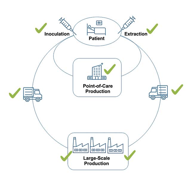
Figure 4. The flow of extracting, producing, and inoculating cells in regenerative medicine; the steps in which quantitative phase imaging may be applied to monitor cell health (check marks). Cells are extracted from the patient and can then be processed either in a smaller facility at the point of care or in a large-scale manufacturing facility. At every step, comprehensive environmental control and cell health monitoring will contribute to a smooth process and reliable cell products, producing successful patient outcomes. Courtesy of Phase Holographic Imaging.
Adapting current QPI technologies to provide continuous cell monitoring will ultimately result in cell-friendly quality control for cell health and differentiation. For example, DHM could be useful for viability control at the cell extraction site, cell health monitoring during the expansion and differentiation steps, and again for cell viability control prior to the reintroduction into the patient. The use of DHM allows vast amounts of data to be generated for each cell sample. In addition, the final cell type verification will need omics analysis — for example, genomics, proteomics, and metabolomics — to ensure the correct gene expression for the type and state of the cells.
A vast amount of data due to these analyses and environmental controls will be generated. It will be a challenge to align cell-monitoring data with environmental and omics data and then organize and present the data comprehensively. The data must be correlated with the individual cell batches and then accompany the cells as they are introduced into the patient. Altogether, data generation, organization, analysis, and storage are all vital parts of the process for successful manufacturing.
Equal to the importance of verifying favorable quality is the ability to predict the outcome of the production process at an early stage. High-quality data will allow for the prediction of the growth and quality of the cells. This will result in more reliable production planning, early indications of deviations in the production process, and more efficient uses of resources.
Currently, an initiative is underway at RegenMed Development Organization, where five companies cooperate in a smart manufacturing project to create a comprehensive, streamlined, controlled manufacturing environment in which cells will be monitored through all necessary steps for expansion and differentiation. QPI will be an important part of the monitoring process, and this project could serve as an impetus for similar integration into future manufacturing.
Meet the authors
Kersti Alm, Ph.D., is the chief scientific officer of Phase Holographic Imaging, a company that develops and manufactures cell analysis tools based on digital holographic imaging; email: [email protected].
Lisa Bodily, M.Sc., is Phase Holographic Imaging’s head of communication; email: [email protected].
References
1. T.L. Nguyen et al. (2022). Quantitative phase imaging: recent advances and expanding potential in biomedicine. ACS Nano, Vol. 16, No. 8, pp. 11516-11544.
2. W. Kim et al. (2022). Recent Advances in Monitoring Stem Cell Status and Differentiation Using Nano-Biosensing Technologies. Nanomaterials, Vol. 12, No. 17, p. 2934.