Miniaturized devices built around various fiber configurations are inserted into the tight, winding systems within the body to guide surgeons at the nanometer level.
Douglas Farmer, Senior Editor
The growing incidence of chronic diseases around the world has driven the expansion of techniques to provide diagnosis at the source, which would ideally involve a minimally invasive — but detail-rich — application of technology. Clinicians are increasingly turning to the endoscope as a solution, and many modern instruments are based on fiber running through their core.
According to Grandview Research, an estimated 225.3 million endoscopic procedures were carried out worldwide in 2022. Prior to the COVID-19 pandemic, an estimated 17.7 million endoscopic procedures were performed annually in the U.S.2
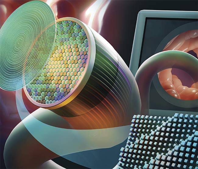
This illustration shows an endoscope, based on meta-optics, that allows for decreased tip length without a reduction in functionality. Adapted with permission from Reference 1.
There are a number of different types of endoscopes, including rigid, flexible, and capsule endoscopes. Rigid endoscopes are characterized by straight, inflexible tubes that facilitate imaging through an opening in the body. Flexible endoscopes integrate fiber optics to allow for maneuverability through various channels in the body. Capsule endoscopes are small devices that can be swallowed and contain a camera and light source to monitor the gastrointestinal system and transmit information to a recording device. The following discussion will focus on flexible endoscopy and the evolving use of optical fiber in diagnostic procedures.
Optical fiber can be made of either glass or plastic, a composition through which light of various wavelengths can easily pass (a process called total internal reflection). The optical fiber generally consists of a core, cladding, and coating and is inherently sensitive to temperature, strain, and pressure. When situated within an endoscope used to explore a patient’s body, it can provide vital data gleaned in vivo from the throat, bladder, and blood vessels (Figure 1).
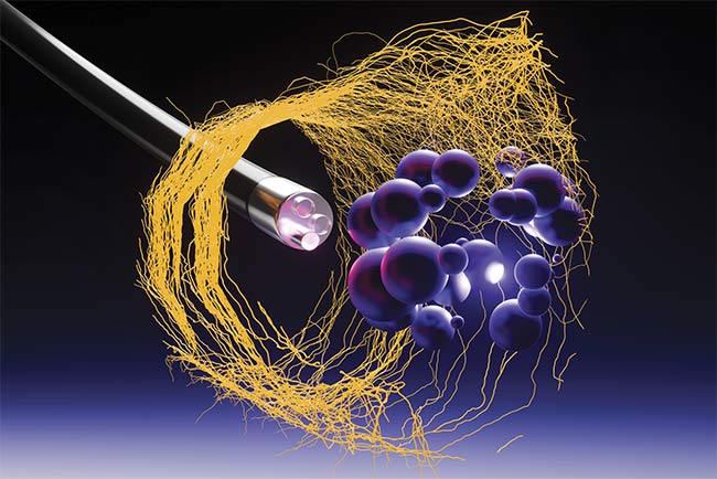
Figure 1. An illustration of an endoscope examining cells in the body. Courtesy of iStock.com/Yuuji.
Singular innovation
Andy Gillooly, business development manager at Fibercore, likens fiber-based endoscopy, in conjunction with other technologies, to a navigation system for diagnostics.
“MRI may be the map, but optical fiber is the GPS system to show where you are during a procedure,” he said.
Endoscopes may be guided by a light pipe, or a bundle of fibers, and are often forward-looking when inserted for minimally invasive diagnostics. Some even take advantage of fiber Bragg gratings (FBGs) at their tips to sense the pressure of resistance, which can warn of blockages targeted for laser ablation in coronary interventions such as angioplasty. The FBGs are inscribed in the coating using an ultraviolet laser.
But as doctors work to precisely define conditions in tight spaces, the challenge becomes how to both deliver light and extract what it reveals with minimal components. At the same time, these miniaturized endoscopic systems must accommodate myriad applications, including optical coherence tomography and laser surgery.
“For example, if you’re looking at the urethra, it is a very small tube and the canula is about the same diameter,” Gillooly said. “In smaller spaces, you can use the fiber itself to achieve endoscopic imaging.”
In single-fiber endoscopy, light is emitted from the fiber and collected back through the cladding with the aid of a piezo mirror, and it is sent to the detector. Fiber can be produced in various sizes. While 125 µm is standard within the telecommunications industry, Fibercore manufactures fiber down to ~50 µm, which is smaller than a human hair and has the advantage of being flexible while easily fitting within standard catheters. Single-fiber endoscopy can be set up in either a forward-facing or side-scanning configuration.
“Not all applications need this capability, and the cost barrier can be considerable,” Gillooly said. “But single fiber can deliver single mode or multimode light. Through manipulation of the fiber, the light beam can come out the side, which when spun can create a stack of an image.”
Increased comfort
And it is about not only enhanced detail but also a more comfortable patient experience. Gillooly recently had an informal conversation about what fiber-based endoscopy can accomplish in this regard with Nilesh Balar, chairman of the department of surgery at Michael’s Medical Center in New Jersey. Balar was pondering the dilemma faced by patients suffering from gastrointestinal distress, such as acid reflux, for which the gold standard is called esophageal manometry. In this procedure, a tube is inserted through the nose and extended into the stomach, with pressure measurements every few centimeters. Generally, patients find this procedure unpleasant, so data can only be extracted for a limited amount of time.
“Manometric measurements are cumbersome but only one or two measurements do not reflect the full picture,” Balar said. “But if you put the fiber across the length of the passage, you could get a clearer idea of how to treat the patient.”
Just as importantly, the 2D images that verify many medical analyses can prove to be inadequate without multiple angles; fiber endoscopes could potentially meet this need, as well.
Single fiber superresolution
Researchers around the world have experimented with the versatility of fiber as a vehicle for imaging in endoscopes. For example, a research team from Zhejiang University in China has been developing a system for superresolution imaging from a thin endoscope through a single, multimode fiber.
Historically, multimode fibers have exhibited extreme sensitivity to bending and other conditions, limiting their transmission capabilities during the examination of tight cavities and vessels within the body. The numerical aperture has also been restricted. But the Zhejiang team successfully demonstrated light-field encoded, subcellular imaging in a single fiber in a technique called spatial-frequency tracking adaptive beacon light-field-encoded endoscopy (STABLE). The instrumentation provides up to 1 kHz disorder tracking frequency, meaning the endoscope is adaptable to bending and other changes. The fiber is integrated into a white light endoscope, enabling cross-scale imaging from microscopic resolution to macroscopic level navigation, and has been tested on pathological samples, lung, and mouse models3.
In their published research, the team gave a detailed description of the system, which was based on a full-vector modulated wavefront that focused distal end reflection light on a single-pixel detector in the spatial frequency domain. This way, the transmission mode is continuously tracked and updated as the fiber’s position changes; the team was able to create 3D images of a sample area targeted for diagnosis. Data collection can be customized, the authors said3.
Qing Yang, a professor of nanophotonics at Zhejiang University in China and corresponding author for the study, said overcoming the resolution limit in tight cavities remains challenging due to the limited numerical aperture determined by small areas and their dynamic surroundings.
“Compared to fiber bundles, single fibers can achieve higher resolution with smaller probe volumes, and are not accompanied by honeycomb noise,” Yang said. “They are more attractive in some narrow-channel imaging scenarios, such as bile and pancreatic ducts or deep brain areas.”
She added single fiber endoscopy is primarily confined to laboratories and not yet widely commercially available.
“We combined the STABLE endoscope with a white light endoscope through the forceps channel, so that it has both a high resolution of multimode fiber and a wide field of view (120 degree field of angle) at the same time,” Yang said.
She said the system must be further developed to include the enlargement of the field of view as well as video imaging speed, which was determined during preclinical studies.
Broad spectra fiber solution
Using super pure material for fiber and its fabrication process enables a broad variety in its spectral range, and the way in which the fiber is produced can also make a difference in its overall use. For example, art photonics GmbH offers fiber set for the broadest spectral range (0.3 to 16 µm), which can accommodate a variety of spectroscopic applications and be integrated into infrared endoscopes and laser catheters to target the specific structure of plaques or tumors.
The polycrystalline infrared (PIR)-fiber is produced by AgCl:AgBr (silver compounds) crystal extrusion through the hard alloy die orifice at very high pressure, using the super-plasticity of this crystal well below its melting point. Slow plasticity extrusion forms unique core/clad nanocrystalline structures of PIR-fibers, flexible and nonbrittle — in sharp contrast with fluoride and chalcogenide infrared glass fibers. The PIR-fiber transmission range is also much broader: 3 to 17 mcm, while ZrF and InF (both fluoride glass) fibers are transparent in 1- to 4-mcm or 1- to 5-mcm ranges and chalcogenide glass fibers in 1- to 6-mcm or 1- to 12-mcm ranges.
As soon as PIR- or infrared glass-fibers are used inside an endoscope, it can perform scanning fiber endoscopy, offering the user MIR vision spectra on top of the common visible and NIR ranges provided by silica glass fibers. Thereby a broader spectral range of imaging can be used when IR fibers can be used with silica fibers.
Viacheslav Artyushenko, president and founder of art photonics GmbH, said that the company works with a partner to provide silica for silicon-based fibers, as well.
“We purify and grow our own crystals, fabricate our core/clad preforms for them, and extrude polycrystalline PIR-fibers with a grain size of 10- to 100-nm scale,” he said. “Since 2020, we’ve also started our own production of hollow glass fibers, which initially were developed by several groups for laser surgery to enable flexible delivery for radiation of CO2 and Er:Yag lasers. Hollow waveguides are glass tubes with reflective coating inside. The hollow waveguide allows the fibers to bend, but they are limited to external surgery operations as their transmission drops fast with bending radius reduction.”
The other type of unique hollow catheters that were previously produced by art photonics were for an order for Eximo Medical from Israel (now AngioDynamics). This construct has worked well in laser angioplasty, during which the double-triple ring of tiny silica fiber guides the laser light to an obstruction, and then the debris resulting from laser ablation is sucked away through a hole in the distal end.
Artyushenko said prior to 2020 his company was partnering with Boston Scientific to produce a single-fiber MIR endoscope, developed by Boston Scientific and composed of PIR-fibers, which was developed to control/prevent overheating of the esophagus wall during an operation on the heart with radio frequency ablation catheters. The project was put on hold.
“The good thing about using MIR information in diagnostics is that all organic molecules have relevant information in this range,” he said.
Endoscopy on the brain
One area of critical clinical need for fiber-based endoscopy is the classification of brain tumors. A team based at Technische Universität (TU) Dresden and at the university hospital Dresden in Germany has been developing autofluorescence-based in vivo endoscopy in the BrainAce project.
Jakob Dremel, a doctoral student in faculty electrical and computer engineering at TU Dresden, said that the typical protocol for diagnosis of a brain tumor is stereotactic biopsy, in which a small needle is inserted into the brain to remove a small amount of tissue. The subsequent chemical staining and interpretation of the data on the sample can take several days to weeks. But Dremel and his fellow researchers conceived that by inserting a tiny fiber endoscope into the biopsy needle, a surgeon could relay information at video rate, skipping time-consuming steps and obviating the removal of tissue (Figure 2)4,5.
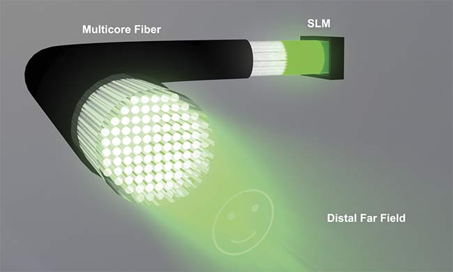
Figure 2. This schematic demonstrates the principle of controlled light-field generation through a 50-cm-long lensless microendoscope. Adapted with permission from Reference 4.
The endoscope they constructed is <1-mm-wide, through which a Fujikura-manufactured coherent fiber bundle (CFB) runs, and a lens and prism projects to the distal end of the fiber. While the CFB produces honeycomb artifacts, Dremel explained that they could be accounted for — and removed from relevant images — with the aid of a cascaded neural network4,5.
“We successfully verified the idea first with a computer simulation, and then with a real setup where a 473-nm laser was used for excitation of the autofluorescence of cryo sections, which was sent through the CFB to the camera,” Dremel said.
The team found that their system achieved 95% accuracy in determination between glioblastoma and metastasis.
The endoscope project is happening in conjunction with an interdepartmental team looking to advance digital holography in diagnostics (Figure 3). The system, called Holoscope, projects computer-generated holograms through the multicore fiber.
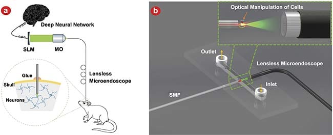
Figure 3. An application of the deep learning enhanced lensless microendoscope. The sketch illustrates employing the device for holographic photoactivation in the mouse brain with minimal invasiveness. Adapted with permission from Reference 4.
“Ordinarily, the fiber bundles can transmit the intensity information but not the phase information,” said Robert Kuschmierz, group leader at the measurement and sensor system techniques at TU Dresden, led by professor Jürgen Czarske. “We wanted to see if we could preserve the phase information in the fibers and correct the transfer function with the aid of an SLM [spatial light modulator] or 3D-printed diffractive optics.”
The team envisions this setup potentially being used for optogenetics in the future, either activating or deactivating specific nerve cells. But first, it could fuel a dramatic improvement of the diagnostics for life-threatening conditions.
“When you’re looking at something like a brain tumor, preoperative data is used, and the CT and MRI images are kept in frame for the procedure,” Dremel said. “But by inserting a fiber optic endoscope, the surgeon can look right into the tissue to make an informed decision. The diagnosis could happen right on the spot and give the surgery a much greater chance of success.”
References
1. J. Froch et al. (2023). Real time full-color imaging in a Meta-optical fiber endoscope. eLight, Vol. 3, No. 13.
2. J. Marroquín-Reyes et al. (2020). National survey regarding the timing of endoscopic procedures during the COVID-19 pandemic. Surg Endosc, Vol. 36, No. 1, pp. 361-366.
3. Z. Wen et al. (2023). Single multimode fibre for in vivo light-field-encoded endoscopic imaging. Nature Photonics, Vol. 17, pp. 679-687.
4. J. Sun et al. (2022). Real-time complex light field generation through a multi-core fiber with deep learning. Sci Rep, Vol. 12, No. 7732.
5. E. Scharf et al. (2020). Video-rate lensless endoscope with self-calibration using wavefront shaping. Opt Lett, Vol. 45,
No. 13, pp. 3629-3632.
Meta-Optics Improves the Scope of Fiber
A team from the University of Washington has developed a fiber-based endoscope that incorporates meta-optics into the design to overcome the challenges associated with coherent fiber bundles and miniaturized components. The team’s collaboration began when Eric Seibel, a research professor in mechanical engineering, attempted to design a scanning fiber endoscope with a miniaturized tip. Decreasing the rigid tip length — which houses the necessary optics — is a vital step in producing an endoscope that is maneuverable and does not damage surrounding tissue.
Their creation was a device derived from a 750-nm-thick silicon nitride (SiN) film and a 500-µm-thick quartz layer and polymer layer, and a 200-µm core fiber. The system allows an ~2.5-mm rigid tip length with a depth of field >30 mm and ~4-mm field of view (opening image). They dubbed their design MOFIE, for meta-optical fiber endoscope1.
Meta-optics are nanoscale diffractive optical elements that shape the phase, amplitude, and spectral response of a wavefront. Historically, however, they have produced aberrations that affect image quality.
“In our system, we used the optics to focus on the sensor, and my task was to integrate diffractive optics with the coherent fiber bundle, which can provide full-color imaging,” said Johannes Fr?ch, a postdoctoral scholar in the department of physics at the University of Washington. “Every lens has aberrations, but we’ve shown that meta-optics can be used to overcome those aberrations.”
The team was able to co-opt computational software with the optics, based on a multichromatic, modulation transfer function — a strategy that helped encode the spatial information into the spectral data collected. This resulted in enhanced imaging from a limited number of data points, Fr?ch said.
To test their system, they placed an organic light-emitting diode screen in front of the meta-optic at the proximal end of the fiber bundle, with an objective, tube lens, and camera at the distal end. The screen was moved to various distances away from the optics. A series of color pictures displayed on the screen were successfully collected by the endoscope.
“We have been collaborating with Luis Savastano, a vascular and endovascular neurosurgeon at University of California San Francisco Health,” he said. “If we can decrease the diameter of endoscopes to just a few tens of microns with single-mode fiber, this impacts how it can be used in that type of application.”
Reference
1. J. Froch et al. (2023). Real time full-color imaging in a Meta-optical fiber endoscope. eLight, Vol. 3, No. 13.