Photonics HandbookBioScan
Technique Enables In Vivo Vascular Imaging at Single-Cell Resolution
A two-photon fluorescence imaging method developed by researchers at the University of Hong Kong and the University of California, Berkeley was able to image the flow of individual blood cells at 1000 2D frames and 1,000,000 line-scans per second in the brains of awake mice.
Such rapid measurements go beyond the reach of conventional two-photon microscopy methods to enable the study of blood flow at the single-cell level in large blood vessels. The ultrafast two-photon fluorescence imaging technique could help scientists better understand how energy is distributed and regulated in both healthy and diseased brains, by revealing blood flow changes at the level of individual blood vessels and across the larger vessel network within the brain.
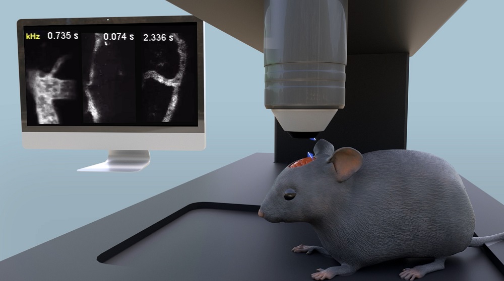
An illustration of cerebral blood flow imaging of an awake mouse using the FACED microscope. Courtesy of the University of Hong Kong.
The researchers used an ultrafast, all-optical scanning method based on free-space angular chirp enhanced delay (FACED). With the line-scanning capability of the FACED module incorporated into a two-photon fluorescence microscope, they were able to demonstrate 1-MHz line scanning and 1-kHz full-frame recording of hemodynamics in the mouse cortex down to 820 µm in depth, at cellular resolution, in vivo.
This level of temporal resolution, which is orders of magnitude greater than existing methods for hemodynamic imaging, enabled the researchers to measure cerebral blood flow up to 49 mm/s.
By directly visualizing red blood cell flow through vessels down to more than 800 µm in depth, the researchers were able to characterize cortical layer-dependent flow velocity distributions of capillaries, obtain radial velocity profiles and 2D kHz velocity mapping of multifile blood flow, and perform red blood cell flux measurements from penetrating blood vessels.
Due to a slow focus scanning mechanism, conventional two-photon fluorescence microscopy is typically limited to a low-flow speed regimen in anesthetized animals. In contrast, the two-photon imaging technology with ultrafast scanning can capture fast-moving flows of red blood cells, even when the animals are awake and have some freedom of movement. In images derived from conventional low-flow speed technology, the blood flow resembles a blur and smear. Images from the ultrafast two-photon fluorescence technology, in comparison, are detailed and clearly show individual cells in fast motion.
“One of the key limitations in other brain imaging technologies has been the resolution of images,” said Kevin Tsia, a professor at the University of Hong Kong. “Our technology is able to deal with this with very fast-moving images, at least 100 times faster than current state-of-the-art options, and capture them down to the individual blood cells. You can count the cells and trace their trajectories.”
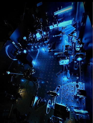
Typical configuration of a FACED imaging system. Courtesy of the University of Hong Kong.
The researchers first deployed a version of their high-speed two-photon fluorescence technology in 2020 to record the electrical signals of neurons in awake mice. Tsia’s team has also applied the technology to cancer screening through the imaging of cancer cells in the blood.
“The work on imaging individual blood cells in vivo is an extension of that work and shows that the technology can be extended to other kinds of neuroscience research, especially cerebral hemodynamics,” Tsia said.
Characterizing blood flow dynamics in vivo is critical to scientists’ understanding of vascular function. The flow of red blood cells is an important indicator of brain activity, which is fueled by energy from blood supply.
Given the versatility of the system in probing many aspects of hemodynamics, from flow velocity, vessel morphology, and velocity profile, to cell counting in bifurcations and penetrating blood vessels, the researchers expect it will be widely used for investigating hemodynamics in living organs.
The research was published in PNAS (www.doi.org/10.1073/pnas.2117346119).
Published: September 2022