Microlenses, imaging chips, and other components have enabled hand-held devices to function in ways previously only possible with laboratory equipment.
FAROOQ AHMED, CONTRIBUTING EDITOR
Spurred by Moore’s law, the principle that the number of transistors on a microchip doubles every two years, the miniaturization of electronic components has resulted in optical devices shrinking as well. While etendue (a characteristic of light and how it diffuses) prevents the exponential size reduction of components, engineers have shrunk optical devices by combining materials and leveraging computing power now available on any smartphone. This integration has also allowed companies and research labs to produce complex devices at low costs, democratizing access to such devices for scientists and nonscientists alike.

Dandelion pollen grains over a wet microscope ruler (10 µm each division) captured using a DIPLE Black objective lens (150× magnification with a
resolution of about 0.7 µm) and an iPhone X. Courtesy of SmartMicroOptics Srl.
Microscopy, among other technologies, has benefitted from these advancements. Lens kits can now turn phones and other hand-held devices into powerful microscopes, and students around the world can use portable microscopes to explore their surroundings. Conventional bright-field microscopy, however, is not the only modality becoming increasingly available thanks to miniaturization: Techniques such as dark-field and holographic imaging, which provide detailed structural information in a label-free manner, are also making inroads — into the palms of users’ hands.
Hand-held bright field
To make scientific devices more readily available, engineers have augmented phones and tablets with microlenses, stages, and companion apps to leverage the devices’ cameras and CMOS imaging chips.
Andrea Antonini, founder and CEO of Genoa, Italy’s SmartMicroOptics Srl, believes these devices will have a lasting impact. “Now, enthusiasm for the devices is growing within the niche of science fans,” he said. “But the technology keeps improving. For the general public, but especially for underserved populations, there will be huge advantages.”
With its kit called DIPLE (Figure 1), SmartMicroOptics enhanced the resolution of smartphone-based microscopy to 0.7 µm, or about 1000× magnification. In the fall of 2019 — through the crowdfunding platform Kickstarter, with which the company had experienced previous success — it quickly raised more than 6× its initial target to fund development.

Figure 1. Genoa, Italy’s SmartMicroOptics developed a portable microscopy kit called DIPLE that leverages smartphone cameras and
processors to capture and analyze images.
Courtesy of SmartMicroOptics Srl.
DIPLE has been in the works for several years, according to Antonini, a physicist who had previously started a photovoltaics company. He said he was inspired by working on microendoscopes for neuroscience applications while at the Italian Institute for Technology, working in Tommaso Fellin’s lab, researching optical approaches to brain function.
“I realized that, with proper changes, some of the technologies that I used for research could be exploited to make innovative add-ons for little cameras like those of smartphones and tablets,” he said.
The complete DIPLE kit, which fits in a box smaller than a smartphone would come in, consists of a stage, an LED light source, and three objectives: 35×, 75×, and 150×. The kit uses a dedicated app and the smartphone or tablet screen as a monitor (lead image). DIPLE has been vetted by researchers and physicians in Europe and the U.S. who have used prototypes to diagnose fecal and blood parasites and assess nonalcoholic fatty liver disease lesions1.
Antonini saw traditional microscope objectives as bulky hindrances to their portability. “I worked on developing a totally new system,” he said.
The lenses are fabricated from glass and plastic polymers but are embedded into aluminum tiles — for compactness and durability and also to allow the lenses to be used with various phone types. “We have designed these parts keeping in mind possible evolutions of the systems, like the use of DIPLE with nonstandard slides, or for hemocytometers,” Antonini said.
Interest in the device has come from several sectors. According to Antonini, “The most common needs are related to the medical field and to science education.”
But veterinarians and oil company technicians have also expressed enthusiasm. “They all need a practical and portable system to prescreen samples in the field or for point-of-care diagnostics — preliminary checks that can massively speed up their processes,” Antonini said.
SmartMicroOptics was set to begin delivery of the kits in May, but the
COVID-19 pandemic curtailed distribution. The kits are now scheduled for delivery this fall.
Smartphones are also getting more intensive upgrades. Researchers based at Duke University and the University of Shanghai for Science and Technology, for example, have integrated dual-functional polymer lenses into a smartphone. These lenses both image and filter light so that the smartphone can be used as a low-cost hand-held fluorescence microscope2. And SmartMicroOptics’ Antonini envisions creating a DIPLE kit to produce dark-field images in smartphones.
Science educators are also taking advantage of portable, low-cost microscopy. To serve educators’ needs and help solve an accessibility problem in science, a group based at Stanford University created the Foldscope (Figure 2), a $1 microscope that unfolds from sturdy paper embedded with optics, according to its website. More than 1 million Foldscopes are in use in over 150 countries, primarily in schools and other educational institutions, and the Foldscope’s resolution of
2 µm (140× magnification) is comparable to that of a classroom microscope3.
Figure 2. A microscope made of paper, the Foldscope, is helping to solve an accessibility problem in science throughout the world. Courtesy of Foldscope Instruments.
The researchers behind the Foldscope recently developed a portable microscopy platform called Octopi that uses correlated bright-field and fluorescence imaging, combined with an automated slide-scanning platform and trained AI, to detect malaria parasites in blood smears. It is powered by a smartphone charger. The Octopi was made for use in rural clinics, to replace the manual microscopy methods currently used in developing countries to detect Plasmodium falciparum parasites in the bloodstreams of patients4. Malaria kills nearly half a million people each year.
Dark field on a chip
Bright-field microscopy is not the only imaging modality to benefit from miniaturization. A group at MIT — led by Mathias Kolle, along with colleagues at Imperial College London and Germany’s Papierfabrik Louisenthal GmbH — developed a postage stamp-size chip that extends dark-field capabilities to conventional microscopes. The chip consists of three layers — gold-film mirrors, quantum dot light sources, and Bragg reflectors, which only allow light at specific incidences to pass through (Figure 3).
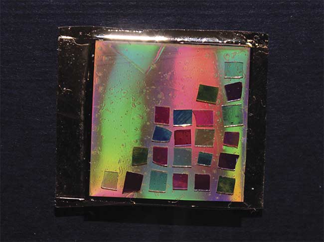
Figure 3. MIT’s Mathias Kolle developed a postage stamp-size chip that extends dark-field capabilities to conventional microscopes. The Bragg mirrors (small squares) generate various angular emission profiles. Courtesy of Cecile Chazot/MIT.
“You can add [our chip] to a cheap, high school-level microscope or even a hand-held, on-chip one,” Kolle said.
Dark-field imaging typically uses filter cubes to block most of the light that enters a microscope’s objective and allows only light diffracted by a sample to pass through. The resultant images appear on a dark background that highlights details and fine structures often washed out by directly transmitted light. Dark-field microscopy is especially useful for visualizing unlabeled subjects, including biological specimens, crystalline structures, and industrial materials.
Like SmartMicroOptics’ Antonini, Kolle saw the drawbacks to traditional dark-field microscopy. “The filters and objectives are bulky and expensive and aren’t suitable to take outside the lab. This means that a lot of people cannot use this functionality,” he said.
In a recent demonstration, the MIT researchers placed their chip on a standard bright-field microscope and produced dark-field images of microbes in seawater taken from Boston Harbor (Figure 4), as well as strains of Escherichia coli bacteria that are difficult to image5.
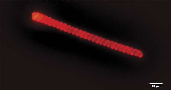
Figure 4. A dark-field image of a 110-µm-long marine microalgae organism observed in a droplet of Boston Harbor seawater. Courtesy of Mathias Kolle/MIT.
Kolle, an associate professor of mechanical engineering at MIT, never intended to change how microscopy is practiced. The development of the dark-field chip began, as it were, organically. He has been studying biological photonic structures for nearly two decades, including how butterfly wings produce their complex and often iridescent colors6. “We were working to simplify these structures and make them more efficient,” Kolle said. “Could we add functionality to them? Could they emit light and even lase?”
Kolle’s goal was to create a chip with tunable microscale lasing capabilities. The project, he admitted, never got to the lasing point.
“But there is a bit of serendipity in this game,” Kolle said. When he and his group studied the emission profile of the butterfly-inspired structures, they saw that although the structures didn’t achieve coherent emissions, the Bragg reflector setup filtered out directly transmitted light in a manner similar to dark-field filter cubes.
Kolle believes that miniaturization will broaden microscopy’s reach. “Part of my group’s research is in that direction,” he said. “The extended size of the optical train in standard microscopes can be a considerable constraint. Miniaturized affordable microscopy systems readily offer opportunities in scenarios where bulky microscopes are out of the question.”
To push miniaturization, he is also working with fluid microlenses that have tunable focal lengths. “These could be used in portable, rugged microscopes,” Kolle said. “At some point, these technologies will merge, and I think you’ll be able to pull out your cellphone, and it will have multifunctional microscopic imaging capabilities.”
Hand-held holography
Perhaps no imaging modality takes advantage of the benefits of miniaturization and the increase in computing power like lensless holographic microscopy. Aydogan Ozcan, a UCLA professor of electrical and computer engineering and a pioneer in small-form biomedical devices, developed several versions of such devices in the last few years7,8. Samples are placed directly atop the image sensor, illuminated by an LED light source, and an algorithm reconstructs and analyzes the image data. “Holography is a computational imaging technique, and today’s most sophisticated computational image reconstruction frameworks exploit deep neural nets,” Ozcan said.
His group tested the microscope by imaging algae in water samples from Los Angeles beaches, as well as airborne pollen and mold spores (Figure 5)9. “This would be useful, for example, for inspectors to sample a part of a building without having to send the acquired sample back to a microbiology lab for further inspection,” he said.
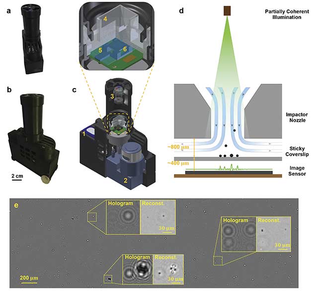
Figure 5. UCLA’s Aydogan Ozcan developed a mobile air-quality detection system that uses deep learning and holography to quantify the density and size of particulate matter, as well as allergens such as pollen particles and mold spores. Photos of the c-Air device from two perspectives (a, b). A quarter is placed beside the device for scale (b). A 3D computer-aided design (CAD) drawing overview of the device (c) — including rechargeable battery (1); vacuum pump (13 L/min) (2); illumination module with fiber-coupled LEDs of red (624 nm), green (527 nm), and blue (470 nm) (3); impaction-based air sampler (4) with a sticky coverslip (5) on top of the image sensor (6). A schematic drawing of impaction-based air sampler on a chip (d). A whole field-of-view differential hologram image of an aerosol sample during sampling, and zoomed-in regions of its reconstruction (e). The device is powered by a rechargeable battery (c1) and controlled by a microcontroller (Raspberry Pi A+). The assembly and packaging are 3D-printed. Courtesy of Ozcan Lab/UCLA.
The group has also developed a hand-held holographic microscope based on this device to detect bacteria and viruses. “You just have to change the image reconstruction engine,” he said. Ozcan hails the merging of these technologies as a “new revolution in data-driven image reconstruction and enhancement methods.”
“Nearly all field measurement tools at the micro- and nanoscales will benefit from the broad availability of these devices,” he said.
SmartMicroOptics’ Antonini concurs: “Point-of-care medical tests aided by portable microscopes will eventually become fundamental tools for better and diffused community medicine, which will reduce the overall costs and improve the efficiency of the health care system.”
www.linkedin.com/in/farooqtheahmed
Acknowledgments
The author would like to thank Andrea Antonini, SmartMicroOptics; Mathias Kolle, MIT; YongKeun Park, Korea Advanced Institute of Science and Technology; and Aydogan Ozcan, UCLA.
References
1. M. Cesaretti et al. (2020). Accurate assessment of nonalcoholic fatty liver disease lesions in liver allograft biopsies by a smartphone platform: a proof of concept. Microsc Res Techniq, Vol. 83, Issue 9, pp. 1025-1031.
2. B. Dai et al. (2019). Colour compound lenses for a portable fluorescence microscope. Light Sci Appl, Vol. 8, article 75.
3. J.S. Cybulski et al. (2014). Foldscope: origami-based paper microscope. PLOS ONE 9(6), e98781.
4. Li H. et al. (2019). Octopi: open configurable high-throughput imaging platform for infectious disease diagnosis in the field. bioRxiv, 684423.
5. C.A.C. Chazot (2020). Luminescent surfaces with tailored angular emission for compact dark-field imaging devices. Nat Photonics, Vol. 14, pp. 310-315.
6. M. Kolle (2011). Photonic Structures Inspired by Nature. Berlin: Springer Science+Business Media.
7. K. de Haan et al. (2019). Deep-learning-based image reconstruction and enhancement in optical microscopy. P IEEE, Vol. 108, No. 1, pp. 30-50.
8. E. McLeod and A. Ozcan (2016). Unconventional methods of imaging: computational microscopy and compact implementations. Rep Prog Phys, Vol. 79, No. 7, p. 076001.
9. Y. Wu et al. (2018). Label-free bioaerosol sensing using mobile microscopy and deep learning. ACS Photonics, Vol. 4, Issue 11, pp. 4617-4627.