FAROOQ AHMED, CONTRIBUTING EDITOR
Spurred by the passage of state legislation last fall, the Southern California Coastal Water Research Project issued a methodology for monitoring microplastics in drinking water. The California regulation was the first of its kind in the U.S. to address widespread microplastic pollution in the environment. In response, an international group of scientists came together to develop guidelines for implementing the regulation, and they identified Raman spectroscopy as a key technique for the chemical identification of microplastics in water.
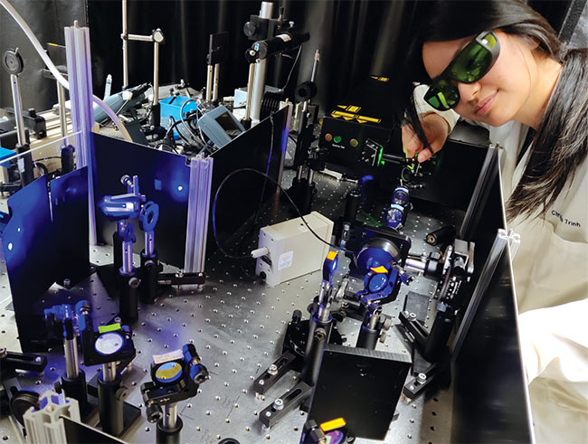
University of California, San Diego graduate student Christina Trinh uses UV resonance Raman spectroscopy to analyze unnatural amino acids in an electron transfer protein. Courtesy of University of California, San Diego/Judy Kim.
Among the reasons for this determination, said Andrew Whitley, who advised on the project, is that, compared to alternative methods such as infrared spectroscopy, Raman spectroscopy allows analysis of much smaller plastic particles while controlling for background interference.
“Raman is winning because of the diffraction limits of other techniques,” said Whitley, who is vice president of global business development at instrument manufacturer HORIBA. Whitley also serves as president of the Society for Applied Spectroscopy.
“It’s amazing what you can do with Raman systems these days,” he said.
Raman spectroscopy is based on the inelastic scattering of light caused by a molecule’s chemical bonds. Detection of this light provides information on the
vibrational energy in the bonds, which carries distinct chemical signatures that can provide information on surface structure as well as other details.
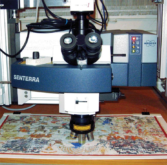
A Raman microscope is mounted on a z stage to enable it to analyze larger works of art. Raman spectroscopy’s ability to nondestructively analyze the chemical composition of materials has helped expand its use for confirming the legitimacy of artwork. Courtesy of Bruker.
But the Raman signal itself is very weak compared with the signals derived from absorption and fluorescence spectroscopy.
Modern Raman instruments typically
deploy light from continuous-wave or pulsed lasers at fixed wavelengths, ranging from the UV to the near infrared (NIR), and detect it with sensitive CCDs. The instruments also incorporate filters to suppress background Rayleigh scattering, and often use confocal apertures, wavelength shifting, or other methods to eliminate fluorescence.
Stable instruments
Advancements in laser and detector technologies as well as their underlying manufacturing processes have led to more reliable and stable Raman instruments, said Tom Tague, applications manager for Fourier transform IR (FTIR) and Raman products at Bruker. These advancements have also enabled smaller and more affordable instruments.
The complex cooling systems that Raman CCDs once required, for example, have been replaced by simpler thermoelectrically cooled detectors. High-end Raman devices, he said, may house up to five lasers that can be software selected and calibrated with little to no user input. Mode shifting of the excitation wavelengths can significantly reduce background fluorescence.
“In my opinion, the ability to shift off fluorescence is one of the biggest innovations of the past several decades,” Tague said. “But, for the last 10 years or so, improvements in Raman instrumentation have slowed, with the focus on making the systems reliable and easy to use. What’s relevant now is the sample.”
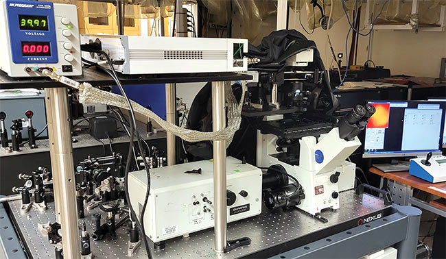
A laser-scanning stimulated Raman spectroscopy (SRS) microscope setup at the University of California, Irvine is powered by a picosecond light source, a lock-in amplifier for isolating the SRS signal, and a computer for collecting imaging data (top). Advanced techniques such as SRS have overcome some of the traditional challenges of spontaneous Raman emission to become new tools with which to monitor cellular processes.
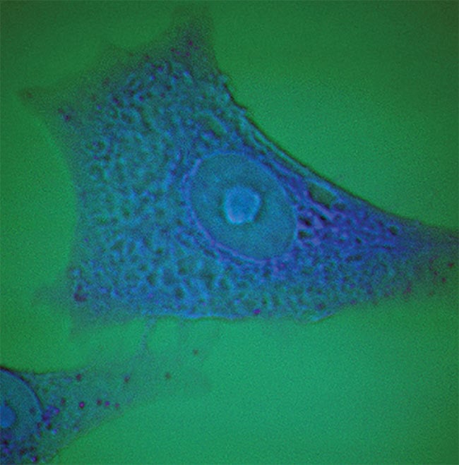
An SRS microscope image of a cultured human lung A549 cell (bottom). The image is segmented into two principal components, which represent the presence of organic matter (blue) and water (green). Courtesy of Courtesy of University of California, San Diego/Judy Kim.
Whitley has seen a change in the industry as well. Where HORIBA once had to educate consumers on why they should use the latest Raman spectrometer, customers are now coming to the company with their ideas.
“Applications are driving the technology,” he said.
Raman evolves
The variety of commercial instruments available as well as the different techniques that can be employed have expanded Raman spectroscopy from a method used primarily by specialists to one with wide applicability, said Judy Kim, a professor of chemistry and biochemistry at the University of California, San Diego.
Techniques such as resonance Raman, SRS, and SERS derive stronger signals from Raman emissions.
According to Kim, there are many benefits to using Raman spectroscopy that nonspecialized users can appreciate. It is nonionizing and label-free, which eliminate the need to prep a sample. The technology provides fast results and enables time-resolved studies. Raman instruments for both macro- and microscopic analysis are stable and widely available. And, she added, there is an active community working on ways to make the technology more sensitive and even faster.
Market research underscores the technology’s potential for growth. Sales of Raman instruments are poised to grow by more than $250 million from 2021 to 2026, to reach a global market value of $861 million, according to research firm MarketsandMarkets. The firm further forecasts that hand-held and portable Raman devices will see the largest growth, driven largely by the biomedical and pharmaceutical sectors.
Kim uses UV resonance Raman (UVRR) spectroscopy and Raman microscopy for studies on biological systems, such as tracking the progression of membrane proteins as they fold and insert themselves into a cell’s lipid bilayer membrane. For these studies, she relies on pulsed deep-UV lasers as well as continuous-wave visible lasers.
Other Raman methods — in particular, stimulated Raman spectroscopy (SRS) — have opened the door for more complex studies, Kim noted. SRS techniques select two separate laser beams with an energy difference that is resonant with a specific molecular vibration. The method overcomes some of the traditional challenges of spontaneous Raman emission and has become a more prominent tool with which to monitor cellular processes.
“I’ve seen a lot of advancements in cell-based Raman,” she said. “You can do very good fundamental science: Take snapshots of bonds forming and breaking — actually see the intersection of chemistry, physics, and biology all in the messy environment of a cell.”
The pharmaceutical industry uses Raman-based macroscopic imaging for quality assurance and quality control, due, in part, to Raman’s nondestructive analysis. Because the detector readout of these instruments is fast and their performance excellent, Bruker’s Tague said, the system can use low-power sources that emit energy below thresholds that might alter or damage samples. These systems still achieve high enough signal-to-noise ratio to allow rapid imaging. Such high-volume imaging is also beneficial for the analysis of biological and geological samples, nanomaterials, and semiconductors. It is also expanding adoption of Raman technology in the analysis of artwork and other precious materials.
Tague described an experience in which he traveled to rural Pennsylvania to authenticate possible Jackson Pollock paintings by using hand-held x-ray
fluorescence tools, as well as portable FTIR and Raman spectroscopy devices. Using the instruments, he was able to determine that the pigments in the artworks matched those used by the painter in other works.
“We were able to get high-quality data in a workshop environment in a very trivial way,” he said. “Trivial” because prior to the use of nondestructive imaging techniques such as Raman, curators often had to remove fragments of paint to confirm the provenance of an artwork.
Signal enhancement
Spontaneous Raman emissions have proved to be a versatile source for collecting analytical data, but nonlinear techniques and signal enhancements are allowing for finer resolution.
Eric Potma, a professor of chemistry at the University of California, Irvine, specializes in nonlinear optical imaging, including coherent Raman scattering (CRS) microscopy — a multiphoton technique that dramatically reduces image acquisition times down to subsecond timescales. “Coherent Raman scattering is very beneficial for biological research, as you can visualize molecules directly through their vibrational signatures and see where they’re moving,” he said.
The setup for this technique, which is not widely commercially available, typically requires two pulsed laser beams to activate specific vibrational modes in a sample. Photomultiplier tubes are often favored over CCDs in CRS setups due to the former’s superior speed and sensitivity.
Other techniques, such as resonance Raman, SRS, and surface-enhanced Raman scattering (SERS), allow spectroscopists to derive stronger signals from Raman emissions, often to tease signals out of a noisy environment.
In the SERS technique, for example, samples are typically affixed to roughened metal surfaces that amplify relatively weak Raman signals. The enhancement can be significant enough to allow for single-molecule detection.
Although the underlying physical mechanisms of SERS are still under debate, Potma credits decades of research for stabilizing the technique and ensuring its reliability. “Every year there are additional beautiful substrates and enhancement structures that are better and easier to fabricate,” he said.
A variation on the SERS theme replaces the roughened metal substrate with a metal nanoparticle. Fay Nicolson, a research fellow at the Dana-Farber Cancer Institute and Harvard Medical School,
recently co-authored a review on the design of SERS nanotags for bioimaging applications1. The paper describes a multistep process in which she and her colleagues build gold nanoparticles and functionalize them with small-molecule Raman reporters such as aromatic thiols or organic dyes. The researchers then affix targeting ligands — often antibodies
— to guide the particles to cancer biomarkers of interest. The particles are injected into anesthetized mice and imaged in the NIR with spatially offset Raman spectroscopy (SORS).
The technique enabled the researchers to probe deeper into tissue than conventional Raman methods allow because
it separated the collection optics and excitation laser. NIR wavelengths correspond to the Raman scattering of the nanotag and have the added benefit of lower autofluorescence in tissue.
“All clinical technologies have some limitation, whether that’s cost, radiation exposure, a lack of molecular information, or [a lack of] depth of measurement,” Nicolson said. “But you can develop ways to overcome these limitations, and that’s where SORS may have an advantage. It’s relatively inexpensive, noninvasive, doesn’t use ionizing radiation, provides molecular information, and has better depth of measurement over other Raman techniques.”
Even with surface enhancements, a signal from spontaneous Raman emission generally cannot penetrate past a few millimeters inside a sample. In contrast, the SORS technique can image Raman signals up to 5 cm deep.
The goal, said HORIBA’s Whitley, is theranostics, in which the same nanotag that images a tumor could be used to destroy it as well. “It’s still a long way away, but hopefully we’ll get it into a human surgical environment,” he said.
Acknowledgments
The author would like to thank Andrew Whitley and Li Yan at HORIBA; Tom Tague at Bruker; Judy Kim at the University of California, San Diego; Eric Potma at the University
of California, Irvine; Fay Nicolson at the Dana-Farber Cancer Institute and Harvard Medical School; Bing Zhao at Jilin University; and James Martin at Sotheby’s.
Reference
1. B. Andreiuk et al. (2022). Design and synthesis of gold nanostars-based SERS nanotags for bioimaging applications.
Nanotheranostics, Vol. 6, No. 1, pp. 10-30, www.doi.org/10.7150/ntno.61244.