Dec. 28, 2023
Laser Speckle Imaging
Laser speckle contrast imaging (LSCI) is a biomedical imaging technique that captures the laser speckle pattern produced when coherent light interacts with a sample, such as human tissue, and is scattered towards a camera detector. Changes in laser speckle pattern over time are related to tissue dynamics and can be used to monitor blood flow noninvasively in vivo. Typically, LSCI systems are large benchtop systems composed of expensive CMOS cameras and laser sources. Because these systems are expensive, bulky, and not portable, they are inaccessible in many health care settings, particularly in low-resource settings. Other methods of measuring blood flow, such as laser doppler flowmetry, share similar drawbacks to benchtop LSCI systems. Recently, research groups have developed portable and wearable LSCI systems for robust use in monitoring blood perfusion across many settings. Development of compact and low-cost perfusion monitoring systems has immense clinical potential for noninvasive monitoring of cardiovascular health.
Key Technologies: Laser speckle contrast imaging, lasers
Raman Spectroscopy
Amyloid fibril are protein aggregates associated with many neurodegenerative diseases, including Alzheimer's. However, the relationship between their molecular structures and toxic activities in the brain remain unclear. To address this problem, we are developing chemical imaging tools using Raman spectroscopy to quantitatively assess how amyloid fibrils aberrantly interact with neurons. A critical step toward achieving this goal involves developing Raman methodologies that enable us to determine amyloid molecular structures with high resolution directly in native biological environments. We recently demonstrated the feasibility of this by showing that structural parameters measured by Raman spectroscopy can be used as constraints to guide molecular dynamics simulations of amyloid fibril models. Our results demonstrate that Raman spectroscopy has the potential to determine detailed, three-dimensional structures of amyloid fibrils in a manner that complements gold-standard methods such as solid-state NMR, as well as enable their direct structural characterization in brain tissue.
Key Technologies: Raman spectroscopy
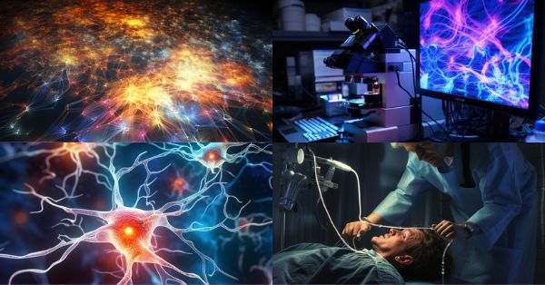
Confocal Microscopy
Although there are various definitions of the physical measurement of detected light, the quantification of "photon number" is the most accurate way to quantify very weak fluorescence light. Therefore, the simplest way of measuring intensity is the number of detected photons. Quantifying fluorescence intensity using a numerical value is very important. Because it is not a device-specific value, this makes it possible to reproduce the same imaging results on different instruments. Furthermore, because the photon number value is universal, researchers can more easily share data with each other without the need to provide experiment-specific parameters. However, there are limitations within confocal microscopy that make it difficult to get consistently accurate photon counts. In this article, we will discuss these limitations and how new detector technologies can help researchers achieve more precise, quality data.
Key Technologies: confocal microscopy
Fiber-Based Endoscopy
The use of fiber in endoscopy allows a minimally invasive approach that can identify problems at their source, whether in the cardiovascular system or the brain. In order to do this, developers have attempted to miniaturize their optics, such as with nanoscale scatter arrays, or eliminating the optics altogether, through measurements drawn directly through the fiber itself. Many endoscopists utilize coherent fiber bundles, which can capture thousands of pixels in a full-color image; by pairing it with a neural network, artifacts can be removed. Other system designers are also turning to a single fiber approach which can be used to scan a sample to extract complicated data. Side-scanning as well as forward-scanning endoscopy is possible, and FBGs can be used to test pressure as guidance for laser ablation. While silica fiber is most common, this limits wavelength detection, so some manufacturers have turned to fiber gleans from crystal extrusion, which is usable in the MIR range and is extremely flexible. The ultimate goal of fiber-based procedures is to allow for real-time diagnostic and surgical information, and for some medical conditions, may eliminate the need for time-consuming biopsy procedures.
Key Technologies: Fiber, coherent fiber bundles, endoscopy, silica fiber, crystal extrusion, FBGs
Download Media Kit